
To take the CE quiz, click here.
This quiz is open until April 2028.
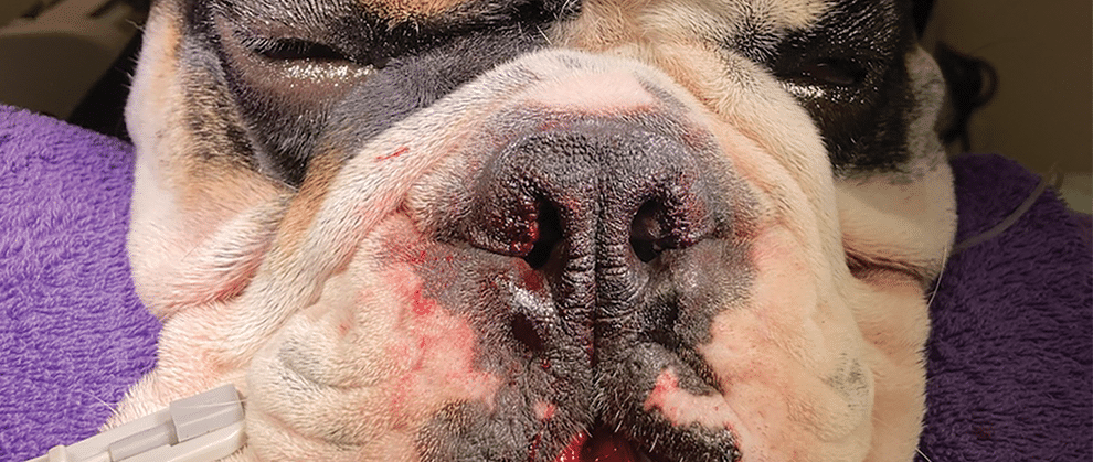
Brachycephalic obstructive airway syndrome (BOAS) affects dogs with brachycephalic skull conformation, such as French and English bulldogs. Clinical signs include stertor, exercise intolerance, and respiratory distress; some dogs also experience gastrointestinal signs (e.g., regurgitation). There are surgical options for correcting some of the components of BOAS (e.g., overlong and thickened soft palate, stenotic nares, everted laryngeal saccules). Standardized perioperative protocols may decrease respiratory complications and/or postoperative regurgitation. Among the variety of surgical procedures for overlong soft palate and stenotic nares, most result in similar positive improvement in respiratory and gastrointestinal signs. Complications can affect up to a quarter of dogs after surgery; most are not life-threatening, although they may result in the need for a temporary tracheostomy or prolonged hospitalization. Postsurgical outcomes are positive for most dogs and result in improved quality of life for the dog and the client.
Take-Home Points
- For most dogs with brachycephalic skull conformation, the benefits from surgical treatment of components of brachycephalic obstructive airway syndrome (BOAS) are improved respiratory and gastrointestinal signs.
- Among dogs undergoing surgery for BOAS component treatment, the risk for respiratory complications is 30 times higher among those in emergent respiratory distress than among those undergoing the surgery on an elective basis, highlighting the value of early intervention.
- Various surgical options exist for treatment of overlong soft palate and stenotic nares; outcomes are similar.
- Standardized protocols for perioperative management of BOAS (e.g., administration of prokinetic agents and proton pump inhibitors, decreased opioid use, preoxygenation before anesthetic induction, use of dexamethasone sodium phosphate) may decrease postoperative regurgitation and/or respiratory complications.
As the popularity of French bulldogs and dogs of other brachycephalic breeds increases, so does the number of dogs presenting to veterinary practices with brachycephalic obstructive airway syndrome (BOAS). Surgical considerations can range from elective for patients with minimal clinical signs to emergent for those exhibiting significant respiratory distress. Understanding when surgery is indicated and the surgeries to recommend can enable more complete client education that leads to better partnering between the veterinarian and the client and improved patient outcomes.
Indications and Preoperative Considerations
Surgical intervention for treatment of BOAS is indicated for dogs with clinical signs associated with BOAS (e.g., stertor, exercise intolerance, respiratory distress). In addition, dogs exhibiting components of BOAS may be candidates for surgical intervention as a preventive measure to decrease the risk for the later sequelae of BOAS (e.g., progressive laryngeal collapse) and future development of clinical signs.
Elective Surgery Cases
When considering surgical interventions for dogs with BOAS, performing thoracic radiography to look for hypoplastic trachea and/or evidence of aspiration pneumonia before inducing general anesthesia is recommended. Dogs with BOAS are at increased risk for aspiration pneumonia due to increased incidence of regurgitation. If aspiration pneumonia is present, delaying elective surgical interventions until the pneumonia has resolved is recommended. For emergent cases with aspiration pneumonia that cannot wait for delayed surgical intervention, aggressive treatment for aspiration pneumonia (e.g., administration of a broad-spectrum antibiotic, nebulization and coupage, supplemental oxygen administration) should be initiated, and clients should be advised that the dog is at increased risk for complications.1 In addition, the overall systemic health of all dogs with BOAS should be evaluated by obtaining a CBC and biochemistry panel before inducing general anesthesia.
Emergent Surgery Cases
For dogs that are in respiratory distress and in need of surgical intervention, oxygen should be provided via an oxygen cage or flow-by oxygen. Placement of an intravenous catheter and administration of sedative medications (e.g., butorphanol, acepromazine) can help decrease distress. Cooling measures may be needed if the dog’s body temperature is elevated. If the provided oxygen support and sedation are not resulting in improved respiration, intubation should be considered after as-needed administration of an intravenous induction agent. Depending on the ventilatory status of the dog, manual ventilation may be necessary.
For some dogs, surgical intervention may be required as soon as the dog is stable. Determining whether upper airway obstruction is the cause of the airway distress can be challenging. Other factors contributing to respiratory distress may be identified by thorough physical examination, upper airway examination, and thoracic radiography. Because of the upper airway swelling and edema that accompany respiratory distress events in brachycephalic dogs, some may require a temporary tracheostomy before or after surgery to allow the swelling to subside. Swelling typically resolves in 2 to 3 days after the distress event, enabling safe removal of the tracheostomy tube; however, dogs should be carefully monitored for reobstruction after tracheostomy tube removal.
Anesthesia Considerations
Compared with nonbrachycephalic dogs, brachycephalic dogs are at an increased risk for complications during and after surgery. One study showed that among brachycephalic dogs, the risk for intra-anesthesia complications is 1.5 times higher and for postanesthesia complications is 4.3 times higher.2 Those risks are commonly related to upper airway obstruction often associated with swelling and perioperative regurgitation and vomiting. Clinicians may individualize decisions for each patient or they may follow standardized protocols before, during, and after anesthesia to decrease the risk.
One method for reducing complications in brachycephalic dogs is preoxygenation before anesthesia induction. Hypoxemia at the time of anesthesia induction may affect any dog, but the risk may be higher for brachycephalic dogs due to the possibility of respiratory obstruction at the time of induction. It has been shown that preoxygenation for 3 minutes before anesthesia induction significantly prolongs time to oxygen desaturation.3
Some standardized protocols combine administration of prokinetic medications (e.g., metoclopramide, cisapride) and a proton pump inhibitor or famotidine preoperatively and encourage intraoperative use of dexamethasone sodium phosphate (dexamethasone-SP) along with perioperatively reduced opioid use to decrease perioperative regurgitation, airway swelling, and aspiration pneumonia. One study reported that use of this protocol significantly decreased postoperative regurgitation, although the number of patients experiencing postoperative pneumonia and respiratory distress did not differ between patients that received the standardized protocol and those that did not.4 Another study reported a significant reduction in postoperative respiratory distress but no changes in postoperative regurgitation or aspiration pneumonia.5 More research on the use of medications alone or in combination should help determine the most ideal protocol to decrease complications.
Another anesthesia goal is to reduce intraoperative and postoperative use of systemic opioids, which may decrease perioperative regurgitation and vomiting leading to aspiration pneumonia. One method for decreasing opioid use while addressing pain control in dogs undergoing surgical treatment of BOAS components is a bilateral maxillary nerve block. A block with lidocaine or levobupivacaine significantly decreases the amount of intraoperative opioids and intraoperative propofol or alfaxalone needed to maintain an appropriate anesthetic plane; however, 1 study reported no difference in the need for postoperative opioid doses between patients that did and did not receive the local block.6
Client Communication Before Surgery
Before their dog undergoes BOAS surgery, clients should be informed of the possible surgical interventions and the risks associated with general anesthesia and the surgery. The brachycephalic risk (BRisk) scoring system can be used to give clients a better idea of their dog’s risk for major complications or death after surgical interventions for BOAS (TABLE 1).7 Risks include complications such as airway swelling (resulting in the need for temporary tracheostomy) and aspiration pneumonia, and clients should be prepared for the possibility of prolonged hospitalization after surgery if complications do occur.
In addition, clients should understand that surgical intervention will often improve but not resolve stertor or exercise intolerance. Gastrointestinal signs such as regurgitation may also improve but not completely resolve. Some BOAS sequelae (e.g., laryngeal collapse) may continue to progress, although at a slower rate after surgery. Although the risk for severe complications is low, some dogs may experience severe upper airway obstruction, aspiration pneumonia, and cardiopulmonary arrest, which may result in death after surgery, and clients should be aware of these possible outcomes before deciding to move forward with surgery.

.
Common BOAS Surgical Techniques
The main components of BOAS that are addressed surgically are an overlong and thickened soft palate, stenotic nares, and everted laryngeal saccules, which are typically all addressed as needed for the individual dog during a single anesthesia event.
Presurgical Upper Airway Examination
Before any surgical intervention, the upper airway should be examined at the time of anesthesia induction. It can also be examined without a planned surgical event to follow; however, some dogs may not be stable enough to extubate without surgical intervention. The veterinarian should anticipate this possible outcome and be prepared to perform a temporary tracheostomy or surgical intervention if extubation is not possible. If surgical intervention or placement of a temporary tracheostomy is not possible, postponing the upper airway examination should be considered, especially for elective cases.
Before anesthesia induction, all necessary items should be obtained to ensure that examination can occur efficiently, allowing for swift intubation to decrease the risk for oxygen desaturation. Items recommended to have available during the upper airway assessment include cotton-tipped applicators, tongue depressors, a headlamp or other light source, and a hemostat or other forceps. Long instruments may be needed to perform laryngeal sacculectomy or other surgical procedures and are recommended if surgical intervention is planned.
During the upper airway examination, the soft palate length and thickness, laryngeal structure and function, tonsils, pharynx, and nares should all be examined. Standardized forms can be used to guide the examination and ensure that all areas are evaluated and findings appropriately documented. Doxapram at a 1-mg/kg dose may be given intravenously to stimulate breathing if needed to assess laryngeal function in dogs that are not taking adequate breaths after anesthesia induction.
Overlong Soft Palate Correction
An overlong soft palate can result in laryngeal obstruction and causes many of the clinical signs associated with BOAS in dogs. In addition, if the palate is overly thick, the oropharynx and nasopharynx may be obstructed. When performing surgery to shorten the palate, the tonsils are used as a landmark to determine the appropriate length of the palate. Ideally, the palate is shortened to the level of the caudal one-third of the tonsil (FIGURE 1), which prevents the palate from continuing to be too long (which could allow clinical signs to continue) or too short (which could allow for aspiration of food or liquids into the nasal cavity).
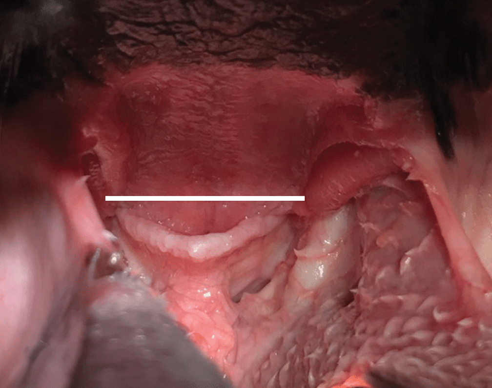
FIGURE 1. Upper airway examination. Appearance of a brachycephalic dog with an overlong soft palate. The white line indicates the level at which the palate should be transected for a standard cut-and-sew staphylectomy.
Staphylectomy
Among the several reported methods for how to perform a staphylectomy in dogs, a commonly used method is a cut-and-sew staphylectomy. For this procedure, the palate is transected at the appropriate level with scissors, and the dorsal and ventral mucosal surfaces are sutured together with 3-0 or 4-0 absorbable suture in a simple continuous pattern.
To manage bleeding from the palate and prevent blood from entering the trachea, the pharynx can be packed caudal to the palate with gauze that is easily retrievable. Also studied for performing a staphylectomy are a variety of other devices (e.g., carbon dioxide laser, monopolar electrocautery, bipolar vessel sealing devices), which generally have been found to be safe and effective with minimal differences noted between outcomes after use of different devices (for more information, see Additional Resources).
Folded-Flap Palatoplasty
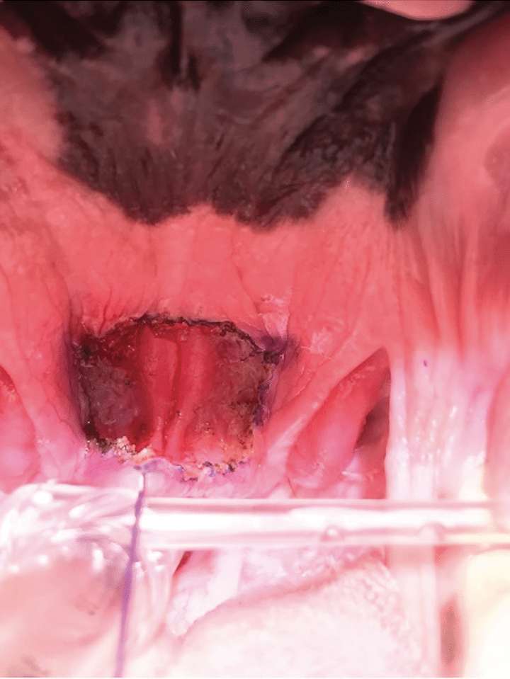
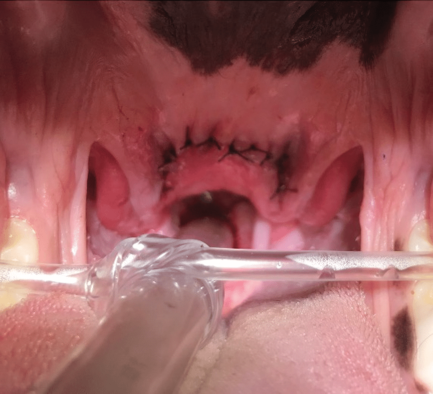
A folded-flap palatoplasty reduces the thickness of the palate while also shortening the palate. The procedure involves excising the ventral mucosa of the palate, the palatinus muscle, and part of the levator veli palatini within a predetermined area on the soft palate, leading to thinning of the palate.8 The caudal-most mucosal surface is advanced rostrally to the cranial-most mucosal surface and the mucosal edges are sutured together with 3-0 to 4-0 absorbable suture in a simple interrupted or simple continuous pattern, leading to shortening of the palate (FIGURE 2).
A study comparing the standard staphylectomy to the folded-flap palatoplasty reported that surgery and anesthesia durations were longer for dogs undergoing the folded-flap palatoplasty than dogs undergoing standard staphylectomy and that anesthetic complications, hospitalization time, and postoperative regurgitation did not differ between the 2 groups.9 Additional research is needed to determine if there are preoperative factors that would indicate which dogs would benefit most from a folded-flap palatoplasty.
Stenotic Nares Correction
Many procedures can be used to widen the nares and decrease airway resistance. One technique is the vertical wedge resection, commonly used because it is a straightforward technique with a good cosmetic outcome. With this procedure, a #11 scalpel blade is used to create a wedge with the open end ventrally on the nasal planum. A deep wedge (pyramid) of tissue is taken and removed, and the epidermis is reapposed with 3-0 to 4-0 absorbable suture in a simple interrupted pattern (FIGURE 3). The surgeon should be sure to remove a deep-enough wedge of tissue as taking only a superficial wedge does not adequately increase the airflow needed to reduce airway resistance. Although a carbon dioxide laser can be used, it can result in temporary depigmentation, which may affect the cosmesis of the procedure. No matter which technique is used, clients should be informed about changes in cosmetic appearance of their dog.


Vertical Wedge, Horizontal Wedge, and Ala Vestibuloplasty
A recent study compared the vertical wedge, horizontal wedge, and ala vestibuloplasty techniques on silicone models.10 Although all 3 procedures resulted in increased cross-sectional area, the ala vestibuloplasty resulted in a significantly larger cross-sectional area. With the ala vestibuloplasty, the axial portion of the alar wing and all of the alar fold are removed. Cosmesis after ala vestibuloplasty is generally very good. Other components seen in dogs with BOAS (e.g., aberrant nasal turbinates) also contribute to nasal airway resistance, and surgical intervention via laser-assisted turbinectomy may be recommended for some dogs that continue to experience respiratory distress after undergoing other surgical interventions.
Everted Laryngeal Saccules Correction
Everted laryngeal saccules represent stage I laryngeal collapse, which is present in many dogs presented for surgical treatment of BOAS components. This author recommends removing everted saccules. Sacculectomy is performed by grasping the tip of the saccule and transecting it at its base, typically with scissors. Damage to the vocal folds during this procedure should be avoided. The procedure can be performed before or after orotracheal intubation. It can be challenging to perform if the dog has laryngeal sensitivity and/or is swallowing and may require administration of additional induction medications. Bleeding is typically stopped by the endotracheal tube. Gentle tissue handling during this procedure will limit upper airway swelling.
Postoperative Management
Dogs should be allowed to slowly wake up from anesthesia and should be extubated when they are as awake as possible but without risking their biting down on the endotracheal tube. Additional endotracheal tubes, intravenous induction agents, sedation, and a laryngoscope should be readily available in case reintubation is necessitated by upper airway obstruction or respiratory distress. Careful monitoring of respiration and oxygenation status after extubation is essential as some dogs may require reintubation in the immediate postoperative period.
If swelling is a concern, phenylephrine or mannitol-dampened gauze may be inserted in the pharynx for 10 to 20 minutes while the dog is still intubated to quickly decrease swelling. After extubation, oxygen support for the first 12 to 24 postsurgical hours may be needed and can be provided via an oxygen cage or nasal oxygen cannula. Dogs are then placed under respiratory watch, in which respiratory rate and effort are checked hourly for the first 12 to 24 hours, depending on how the patient is recovering from general anesthesia.
To prevent regurgitation, a postoperative constant-rate infusion of metoclopramide or administration of cisapride can be considered. A 0.1-mg/kg IV dose of dexamethasone-SP is often given intraoperatively and can be repeated postoperatively as needed to decrease swelling. NSAIDs can also be considered as an alternative to steroids to decrease airway swelling if preferred by the clinician; however, steroids and NSAIDs should not be given together due to the risk for gastrointestinal ulceration.
To decrease swelling of the upper airway during the recovery period, dogs should be kept calm while hospitalized and after discharge. During hospitalization, intravenous sedation (e.g., acepromazine, butorphanol) can be used until the dog is able to take oral medications (e.g., trazodone). Sedation should be used cautiously, avoiding oversedation, which can cause respiratory depression or obstruction if the patient is unable to appropriately position its head to keep the airway open. Trazodone can be used at home for 10 to 14 days after surgery.
To prevent trauma to the healing soft palate in dogs that underwent staphylectomy, patients should be fed only soft food meatballs for the first 2 weeks after surgery. Dogs that underwent any procedure to correct stenotic nares should wear an Elizabethan collar to prevent trauma to the healing incisions.
In hospitals where it is possible, client-assisted recovery and same-day discharge of brachycephalic dogs has gained popularity for decreasing anxiety and possibly complications. One study compared outcomes of hospital-only and client-assisted recovery.11 In the study, 1 group of dogs underwent standard postoperative recovery in the hospital for at least 24 hours, and another group had the client present in the kennel with them during recovery and were sent home when they were breathing normally and able to walk around the recovery room. Complication rates for dogs that underwent client-assisted recovery were only 2% compared with 28% for dogs undergoing standard recovery. Thus, further study may indicate client-assisted recovery as an option after surgery for BOAS.
Postoperative Complications
The most common postoperative complications after BOAS surgery are upper airway obstruction and aspiration pneumonia, which affect up to 23% of dogs.12 Respiratory complications are more common among older dogs, dogs with laryngeal or tracheal collapse, and dogs that require emergency surgical intervention.
Dogs with airway swelling and obstruction not improved by oxygen administration and steroids may need a temporary tracheostomy, left in place 2 to 3 days while the dog is hospitalized to allow time for the swelling to decrease. Another option is nasotracheal intubation to allow for oxygen administration while bypassing the upper airway. With this procedure, a 5- to 10-French tube is inserted through the naris and exteriorized through the oral cavity. The dog is extubated, and the tube is passed into the trachea to the level of the thoracic inlet with oxygen flow delivered through the tube.
One study reported that no dogs with a nasotracheal tube experienced respiratory distress postoperatively and that one-third of the dogs without a nasotracheal tube did experience respiratory distress.13 Concerns regarding nasotracheal intubation include the possibility that the tube could become obstructed, compressed, or dislodged, thereby preventing oxygen delivery. Although not necessary in every dog undergoing BOAS surgery, nasotracheal intubation is an option to avoid a temporary tracheostomy for some dogs experiencing respiratory complications after extubation that are not responsive to flow-by oxygen.
Postoperative regurgitation may be worse than preoperative regurgitation and may be secondary to ileus resulting from general anesthesia or administration of opioid medications during surgical intervention or from upper airway swelling after surgery leading to increased airway resistance and worsening gastrointestinal signs. For some dogs, decreased use of pure µ opioids and enhanced use of prokinetic medications (e.g., metoclopramide, cisapride) may limit regurgitation after surgery. Aspiration pneumonia may develop as a result of postoperative regurgitation. Aspiration pneumonia in those dogs is treated with oxygen support, antibiotics, and nebulization and coupage, as would be recommended for other dogs with pneumonia.
Surgical Outcomes
Surgery for BOAS can significantly improve the quality of life for some dogs, despite the fact that some components or sequelae of BOAS (e.g., macroglossia, hypoplastic trachea) cannot be completely remedied with surgery at this time. One study reported that 71% of dogs undergoing BOAS surgery showed decreased respiratory signs.14 When surveyed, approximately 90% of owners noticed reduced gastrointestinal disorders in their dog after BOAS surgery.15 One of the most notable improvements was decreased incidence of postanesthesia complications at subsequent anesthetic events after BOAS surgery; 1 study reported 79% decreased odds of a complication.16
Summary
BOAS is complex disease that affects many dogs with brachycephalic conformation. Despite risk for postsurgical complications in these dogs, many benefit greatly from the surgery and most exhibit reduced respiratory and gastrointestinal signs. Successful outcomes depend on thorough client communication and strict attention to surgical technique, preoperative management, and postoperative treatment plans.
- Dunié-Mérigot A, Bouvy B, Poncet C. Comparative use of CO2 laser, diode laser and monopolar electrocautery for resection of the soft palate in dogs with brachycephalic airway obstructive syndrome. Vet Rec. 2010;167(18):700-704. doi:10.1136/vr.c5107
- Grubb T. Anesthesia and analgesia in brachycephalic dogs. Todays Vet Pract. 2022;12(5):48-62.
- Kirsch MS, Spector D, Kalafut SR, Moore GE, McDougall R. Comparison of carbon dioxide laser versus bipolar vessel device for staphylectomy for the treatment of brachycephalic obstructive airway syndrome. Can Vet J. 2019;60(2):160-166.
References
- Ree JJ, Milovancev M, Macintyre LA, Townsend KL. Factors associated with major complications in the short-term postoperative period in dogs undergoing surgery for brachycephalic airway syndrome. Can Vet J. 2016;57(9):976-980.
- Gruenheid M, Aarnes TK, McLoughlin MA, et al. Risk of anesthesia-related complications in brachycephalic dogs. JAVMA. 2018;253(3):301-306. doi:10.2460/JAVMA.253.3.301
- McNally EM, Robertson SA, Pablo LS. Comparison of time to desaturation between preoxygenated and nonpreoxygenated dogs following sedation with acepromazine maleate and morphine and induction of anesthesia with propofol. Am J Vet Res. 2009;70(11):1333-1338. doi:10.2460/ajvr.70.11.1333
- Costa RS, Abelson AL, Lindsey JC. Wetmore LA. Postoperative regurgitation and respiratory complications in brachycephalic dogs undergoing airway surgery before and after implementation of a standardized perianesthetic protocol. JAVMA. 2020;256(8):899-905. doi:10.2460/javma.256.8.899
- Hill M, Lepiz ML, Ida KK, Rutter C, Mankin KT, Dickerson V. Implementation of a standard perioperative protocol reduces postoperative respiratory distress events in dogs undergoing surgical correction of brachycephalic obstructive airway syndrome. JAVMA. 2025;263(5):590-598. doi:10.2460/JAVMA.24.09.0598
- De Gennaro C, Vettorato,E, Corletto F. Evaluation of bilateral maxillary nerve block in dogs undergoing surgery for brachycephalic obstructive airway syndrome. Can Vet J. 2022;63(1):67-73.
- Tarricone J, Hayes GM, Singh A, Davis G. Development and validation of a brachycephalic risk (BRisk) score to predict the risk of complications in dogs presenting for surgical treatment of brachycephalic obstructive airway syndrome. Vet Surg. 2019;48(7):1253-1261. doi:10.1111/vsu.13291
- Findji L, Dupré G. Folded flap palatoplasty for treatment of elongated soft palates in 55 dogs. Wien Tierarztl Monatsschr. 2008;95(3):56-63.
- Miller AK, Regier PJ, Colee JC. Complications and outcome following staphylectomy and folded flap palatoplasty in dogs with brachycephalic obstructive airway syndrome. Vet Surg. 2024;53(1):29-37. doi:10.1111/vsu.13994
- Franklin PH, Riggs J, Liu NC. Comparison of the effectiveness of three different rhinoplasty techniques to correct stenotic nostrils using silicone models: a case study. Vet Surg. 2024;53(1):104-112. doi:10.1111/vsu.14041
- Camarasa JJ, Gordo I, Bird FG, Vallefuoco R, Longley M, Brissot HN. Owner-assisted recovery and early discharge after surgical treatment in dogs with brachycephalic obstructive airway syndrome. J Small Anim Pract. 2023;64(11):680-686. doi:10.1111/jsap.13647
- Lindsay B, Cook D, Wetzel J-M, Siess S, Moses P. Brachycephalic airway syndrome: management of post-operative respiratory complications in 248 dogs. Aust Vet J. 2020;98(5):173-180. doi:10.1111/avj.12926
- Senn D, Sigrist N, Forterre F, Howard J, Spreng D. Retrospective evaluation of postoperative nasotracheal tubes for oxygen supplementation in dogs following surgery for brachycephalic syndrome: 36 cases (2003-2007). J Vet Emerg Crit Care (San Antonio). 2011;21(3):261-267. doi:10.1111/j.1476-4431.2011.00612.x
- Seneviratne M, Kaye BM, Ter Haar G. Prognostic indicators of short-term outcome in dogs undergoing surgery for brachycephalic obstructive airway syndrome. Vet Rec. 2020;187(10):403. doi:10.1136/vr.105624
- Poncet CM, Dupre GP, Freiche VG, Bouvy BM. Long-term results of upper respiratory syndrome surgery and gastrointestinal tract medical treatment in 51 brachycephalic dogs. J Small Anim Pract. 2006;47(3):137-142. doi:10.1111/j.1748-5827.2006.00057.x
- Doyle CR, Aarnes TK, Ballash GA, et al. Anesthetic risk during subsequent anesthetic events in brachycephalic dogs that have undergone corrective airway surgery: 45 cases (2007-2019). JAVMA. 2020;257(7):744-749. doi:10.2460/javma.257.7.744
CE Quiz
This article has been submitted for RACE approval for 1 hour of continuing education credit and will be opened for enrollment upon approval. To receive credit, take the test at vetfolio.com. Free registration is required. Questions and answers online may differ from those below. Tests are valid for 2 years from the date of approval.
1. How long should a brachycephalic dog be preoxygenated before anesthesia induction to significantly prolong the time until oxygen desaturation?
a. 1 minute
b. 3 minutes
c. 5 minutes
d. 7 minutes
2. Respiratory complications after surgery to correct components of brachycephalic obstructive airway syndrome (BOAS) are more common among dogs with which of the following conditions?
a. Dogs that are younger
b. Dogs without clinical signs
c. Dogs that undergo emergency surgical intervention
d. Dogs with macroglossia
3. A standardized perioperative protocol including prokinetic medications and proton pump inhibitors or famotidine, encouraging the use of dexamethasone sodium phosphate, and decreasing opioid use may decrease postoperative respiratory distress and/or postoperative regurgitation.
a. True
b. False
4. When performing a staphylectomy for overlong soft palate, at what level should the palate be transected?
a. Caudal one-third of the tonsil
b. Midpoint of the tonsil
c. Cranial one-third of the tonsil
d. Cranial tip of the tonsil
5. Use of a bilateral maxillary nerve block before BOAS surgery results in decreased use of opioids intraoperatively.
a. True
b. False