
To take the CE quiz, click here.
This quiz is open until April 2028.

Brachycephalic obstructive airway syndrome (BOAS) is a progressive and often life-threatening respiratory condition that affects brachycephalic breeds such as French bulldogs, English bulldogs, pugs, and Boston terriers. A mismatch of shortened skull structures and excess soft tissues leads to upper airway obstructions in these breeds. This article reviews the primary anatomic abnormalities, as well as secondary respiratory and gastrointestinal complications, associated with BOAS along with key conformation and lifestyle risk factors that exacerbate the clinical signs of the syndrome. Early recognition, preventive strategies, and long-term reformation of breed standards are essential to reducing the prevalence and severity of BOAS.
Take-Home Points
- Brachycephalic obstructive airway syndrome is a collection of anatomic abnormalities related to the craniofacial conformation of brachycephalic dogs.
- The primary abnormalities of the syndrome are stenotic nares, aberrant nasal turbinates, elongated/hyperplastic soft palate, macroglossia, and hypoplastic trachea.
- These primary abnormalities can lead to secondary abnormalities, which include laryngeal collapse, redundant oropharyngeal tissue, everted tonsils, and gastrointestinal disease.
- In conjunction, primary and secondary abnormalities lead to progressive airway resistance, stertor, exercise intolerance, reduction in quality of life, and potential respiratory obstruction.
- Preventive measures such as selective breeding, early intervention, environment modification, and management of body condition score may help alleviate some of the clinical signs.
Coinciding with the growing popularity of brachycephalic breeds, brachycephalic obstructive airway syndrome (BOAS) has become increasingly relevant to veterinarians over the past several years. French bulldogs, English bulldogs, pugs, and Boston terriers have rapidly gained popularity; according to the American Kennel Club, the French bulldog has been the most popular breed in the United States for 4 consecutive years.1
Because of the growing prevalence of BOAS and its often life-threatening complications, a strong understanding of the syndrome is essential. Dogs with BOAS have a myriad of clinical signs, the most severe of which is respiratory distress, which can progress to complete respiratory obstruction. Familiarity with the key anatomic components of BOAS and their clinical manifestations is crucial to making treatment plans and implementing preventive measures. BOAS is fundamentally characterized by increased airway resistance, and every therapeutic and preventive intervention aims to reduce this resistance and improve airflow.
BOAS is a collection of anatomic abnormalities related to the craniofacial conformation of brachycephalic dogs. The syndrome arises from a mismatch between foreshortened craniofacial bones and the soft tissues within the nasal passages, nasopharynx, and laryngeal region (e.g., palate, tongue, nasal turbinates, mucosa). This mismatch leads to increased airway turbulence and resistance. The 5 primary abnormalities in dogs with BOAS are stenotic nares, aberrant nasal turbinates, elongated/hyperplastic soft palate, relative macroglossia, and hypoplastic trachea (FIGURE 1). These abnormalities can contribute individually to the classic clinical signs of BOAS; in combination, they synergize, leading to potentially life-threatening sequelae.
Additional conformation changes are often present in dogs with BOAS; these include thickened submucosae (i.e., mucosal redundancy) and increased goblet cells. Over time, these abnormalities generate increased negative pressure during inspiration, which ultimately fosters a dynamic laryngeal collapse and/or an eversion of adjacent tissues (e.g., everted laryngeal saccules, everted tonsils). Ultimately, a multilevel upper airway obstruction with increased respiratory effort predisposes these dogs to chronic hypoxia, hypercapnia, and negative intrathoracic pressure. Additional secondary sequelae include changes to the pulmonary and gastrointestinal systems such as gastroesophageal reflux and hiatal herniation. The clinical signs of secondary sequelae can vary widely and correlate closely with the extent of associated anatomic abnormalities.

Figure 1. Cross-section of mesocephalic and brachycephalic dogs demonstrating the shortened skull conformation and excessive soft tissue associated with brachycephaly. Airway resistance is increased at the stenotic nares and by excessive nasal turbinates, contributing to an elongated soft palate and everted laryngeal saccules. These changes may be further exacerbated by relative macroglossia. Illustration: Kip Carter
Primary Abnormalities
Stenotic Nares and Aberrant Nasal Turbinates
Stenotic nares are one of the earliest, most accessible, and most common lesions contributing to airway resistance (FIGURE 2). Research indicates that up to 75% of dogs with BOAS that pursue surgery have stenotic nares.2 Affected dogs have narrowed external nostrils as well as a reduced diameter of the nasal vestibule leading into the nasal cavity. Minimal stenosis of the nares can significantly increase inspiratory load, and even a slight reduction in the cross-sectional area of the nasal vestibule produces exponentially greater airflow resistance. Changes in the nasal cavity account for 80% of the airway resistance in dogs with BOAS.3 In French bulldogs, English bulldogs, and pugs, stenotic nares are a significant predictor of BOAS.4 A 2017 study found that 75% of French bulldogs had moderately to severely stenotic nares compared to 65% of pugs and 44% of English bulldogs.4 Furthermore, dogs with moderately to severely stenotic nares often exhibit immobile nostril wings, which prevent the nares from abducting normally during exercise. This absence of abduction leads to poor thermal regulation and excess negative pressure within the airway.
Abnormally developed or displaced nasal turbinates, referred to as aberrant nasal turbinates, are another frequent malformation in dogs with BOAS. This abnormality results from continued turbinate growth despite cessation of midface growth in young brachycephalic dogs, leading to oversized nasal turbinates that statically impede airflow through the nares.5 Aberrant nasal turbinates increase nasal mucosa contact points and further increase nasal airway resistance and turbulent nasal airflow. Other sequelae include a reduction in sinus ventilation and olfactory function as well as a predisposition for nasal tissue edema and lymphoplasmacytic rhinitis.6,7
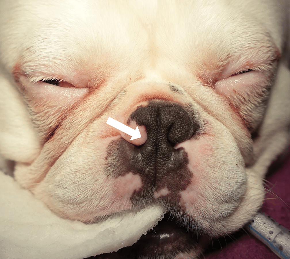
FIGURE 2. A 6-month-old French bulldog under anesthesia for a brachycephalic obstructive airway syndrome procedure. Note the axial displacement of the alar wing (arrow) that markedly reduces the diameter of the external nare.
Elongated Soft Palate and Mucosal Hyperplasia
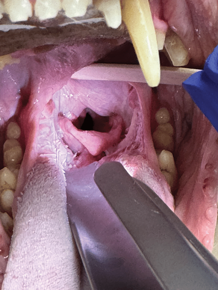
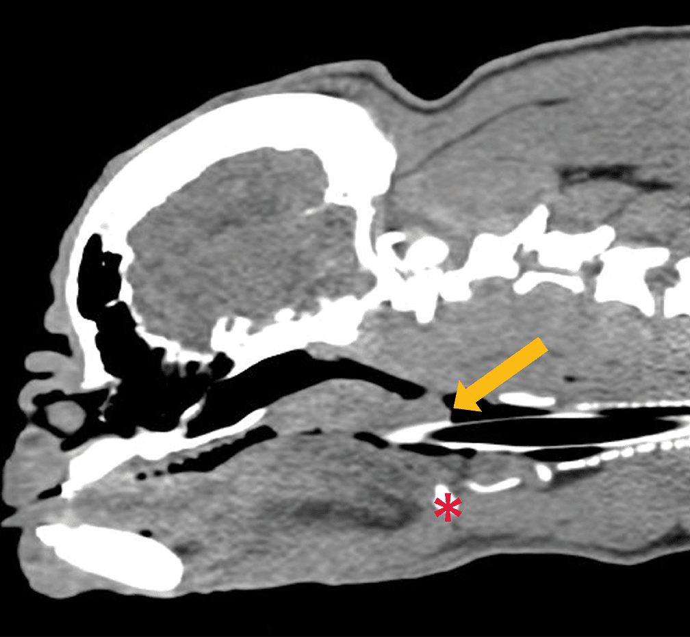
The hallmark lesion of BOAS is an elongated and thickened soft palate. Elongation of the soft palate produces the stertor typical of BOAS, which is the clinical sign most frequently reported by clients. Like many other BOAS-related abnormalities, an elongated soft palate is the result of a disproportionate skull-to-tissue ratio. An enlarged soft palate can lead to laryngeal, oropharyngeal, and nasopharyngeal obstruction, which can severely worsen clinical signs and increase the risk for complete upper airway obstruction.2 Additionally, an elongated soft palate can overlap the epiglottis, which can cause retching and gagging (FIGURES 3 AND 4). A sedated airway examination is necessary to assess the soft palate. Histologically, mucous gland hyperplasia and edema are most notable in elongated soft palates; these findings are accompanied by reduced muscle mass resulting from necrosis and degeneration. (These histologic changes are similar to those observed in humans with obstructive sleep apnea syndrome.8,9) In most patients, the conditions associated with an elongated soft palate worsen over time due to chronically increased airway resistance and altered pressure dynamics.
Relative Macroglossia
Macroglossia refers to an enlarged tongue. Dogs predisposed to BOAS typically have relative macroglossia in which the tongue is disproportionately large for the size of the mouth. Compared to mesocephalic breeds, brachycephalic breeds have a greater tongue volume relative to body weight, skull length, and ratio of skull length to width. Due to this relative macroglossia, the ratio of air to soft tissue is decreased by 60% compared with unaffected dogs, which further increases airway resistance.10 Macroglossia causes dorsal displacement of the soft palate, which narrows the nasopharynx and significantly increases airway resistance, thereby impeding airflow (FIGURE 5).11 Pugs have a considerably smaller tongue volume compared to French and English bulldogs, thus the role of macroglossia in this breed is questionable.
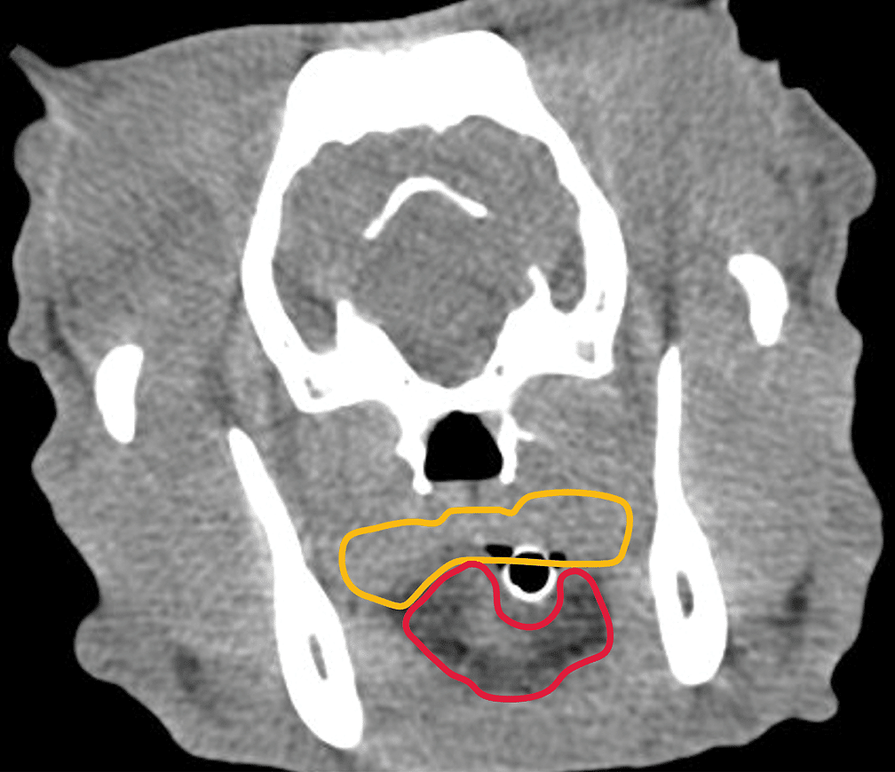
FIGURE 5. A transverse computed tomography image of a 1-year-old French bulldog with an endotracheal tube in place. The area outlined in yellow indicates a thickened soft palate, and the area outlined in red indicates relative macroglossia. Note how they combine to decrease the functional airway volume to the dark area around the endotracheal tube.
Hypoplastic Trachea
A hypoplastic trachea is an underdeveloped or abnormally narrow trachea, typically characterized by small, rigid tracheal cartilage rings and shortened or absent dorsal tracheal membrane. Thoracic radiographs are used to diagnose a dog with hypoplastic trachea (FIGURE 6). Clinicians should use caution when diagnosing this in a juvenile dog as it may improve as the dog reaches skeletal maturity. The most affected breeds are English bulldogs and brachycephalic breeds with screw tails.12 This abnormality is not always associated with clinical signs and is absent in some dogs with BOAS. Nonetheless, this abnormality can exacerbate clinical signs and is a negative prognostic indicator with concurrent bronchopneumonia.12
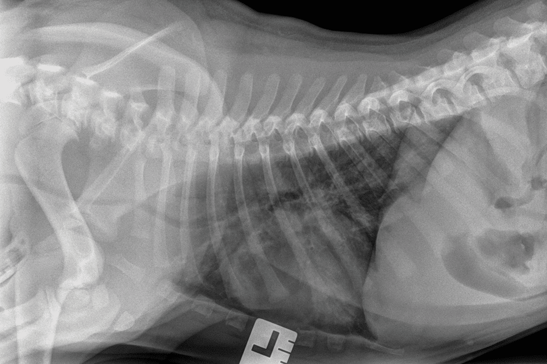
FIGURE 6. A left lateral thoracic radiograph of a 6-month-old English bulldog with suspected hypoplastic trachea.
Secondary Abnormalities
In addition to these primary anatomic abnormalities, dogs with BOAS often develop secondary respiratory changes that worsen airway resistance and increase breathing effort. Chronic negative pressure as well as airway turbulence, irritation, and inflammation commonly lead to edema, hypertrophy, and eversion of the palatine tonsils, further narrowing the pharyngeal space. Enlarged tonsils are associated with respiratory infection and exercise intolerance.13
Over time, laryngeal cartilage fatigue and chondromalacia reduce laryngeal stiffness, predisposing affected dogs to eversion of the laryngeal ventricles (saccules) and progressive laryngeal collapse.14 Laryngeal collapse results from a progressive failure of the laryngeal cartilages, particularly the arytenoid cartilages. Stage I collapse involves eversion of the laryngeal ventricles and accounts for approximately 58% of dogs with BOAS that require surgery (VIDEO 1). These everted laryngeal ventricles, which arise from the mucosal lining of the laryngeal ventricular recesses located rostral to the vocal folds, are drawn into the airway by increased inspiratory pressure, further diminishing airway patency. Stages II and III entail continued structural failure, characterized by medial displacement—and sometimes overlapping—of the cuneiform and corniculate processes, resulting in severe airway narrowing. Stages II and III generally indicate end-stage, irreversible disease.15
Beyond these primary and secondary abnormalities, dogs with BOAS frequently exhibit additional abnormalities such as redundant pharyngeal tissue, bronchial collapse (reported in over 85% of dogs and most often involving the left mainstem and left cranial dorsal bronchi in 1 study),15 hiatal herniation, gastroesophageal reflux, and regurgitation. The complications of BOAS extend beyond the respiratory system because chronic negative intrathoracic pressure leads to gagging, vomiting, and regurgitating, which increases the risk for aspiration pneumonia and further worsens respiratory compromise. Gastrointestinal disease is present in up to 97% of brachycephalic dogs exhibiting respiratory signs.16-18 In addition, these breeds have a larger esophageal hiatus, which predisposes them to reflux, regurgitation, and hiatal herniation.19,20 Some research currently supports that these gastrointestinal changes are primary in nature.20
Risk Factors and Their Clinical Effects
Additional conformation abnormalities contribute to the risk factors and predictive value of BOAS prevalence. An understanding of the abnormalities that are not as commonly discussed may enable the prediction of a patient’s risk level and inform clinical recommendations.
Craniofacial ratio (CFR) is the length of the muzzle divided by the cranial length of the face; a shorter CFR indicates a flatter, more smooshed face. The risk for BOAS increases sharply in a nonlinear manner as muzzle length shortens.11 Although CFR may be a predictor of BOAS, it should not be used as a sole predictor; clinicians should use CFR in conjunction with other conformation attributes and clinical signs.
Neck dimensions, particularly girth and length, have been shown to affect the risk for severe BOAS. In French and English bulldogs and pugs, a neck girth-to-length ratio greater than 0.71 in males predicts BOAS with approximately 70% sensitivity and specificity.4 Similar associations between neck girth-to-length ratio and BOAS have been noted in French bulldogs.
Body condition score (BCS) plays the most clinically significant role in the risk for BOAS. Obesity is a robust risk factor for BOAS in pugs and bulldogs. Dogs with an increased BCS begin to store adipose tissue in tissues that surround the airway (e.g., tongue, palate), further narrowing the airway. One study of brachycephalic dogs observed by primary care veterinarians (i.e., not referred to a specialty clinic) found that 57% of dogs were overweight (BCS > 5/9) and 10% were obese (BCS ≥ 8/9).11
Treatment of BOAS
Diagnosis
The ability to assess, diagnose, and stabilize patients with BOAS is critical for any practitioner. A thorough physical exam, including examination of BCS, should be performed; additional focus should be given to externally observable BOAS features, including assessment of nares, auscultation of the trachea and larynx, and respiratory effort at rest and while walking. Radiography can be a useful diagnostic tool to assess tracheal diameter and detect lung pathology (e.g., aspiration pneumonia) in stable patients. A sedated airway exam enables assessment of an elongated soft palate and/or other obstructive tissue; however, caution should be exercised as severely affected dogs can have difficulty recovering from sedation. A sedated airway exam is not recommended if a clinician is not comfortable or prepared to intubate and either provide surgical intervention or transfer to a qualified facility.
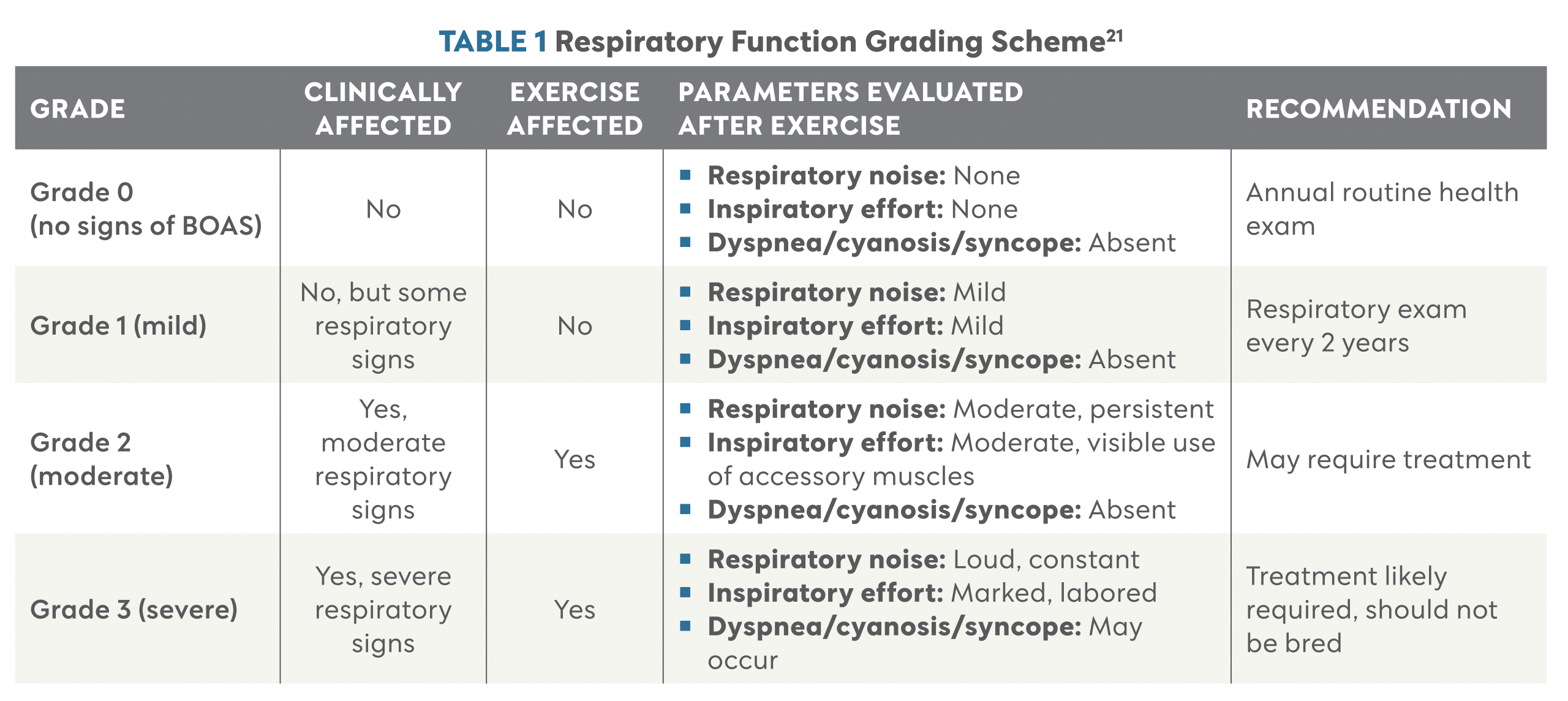
The University of Cambridge, in collaboration with the Royal Kennel Club, has developed a grading scheme for brachycephalic breeds that serves as a reference for the severity of BOAS, provides guidance on when airway exams should be performed, and identifies which animals should not be bred.21 The Respiratory Function Grading Scheme involves grading clinical signs when a dog is calm, then 4 to 5 minutes after moderate exercise. The scheme primarily evaluates 3 categories: respiratory noise, inspiratory effort, and dyspnea/cyanosis/syncope (TABLE 1). The grading scheme ranges from 0 through 3, with 3 being the most severely affected. Additional recommendations include reassessing dogs with grade 1 every 2 years and refraining from breeding dogs with grade 3. The University of Cambridge website provides an in-depth breakdown of the recommendations using this grading scheme.21
Triage
Dogs with BOAS often arrive in respiratory distress; therefore, preparedness for prompt and appropriate stabilization is essential. Dogs with increased respiratory effort should receive supplemental oxygen via flow-by or mask as tolerated. If a dog is hyperthermic, active cooling measures, including ventilation with cool air and application of cold or wet towels, should be initiated. Intravenous fluid therapy should be considered to support thermoregulation and perfusion. Some patients may benefit from mild sedation to reduce anxiety and respiratory effort, provided it can be administered safely. If respiratory effort does not improve within approximately 10 minutes of oxygen supplementation, cooling, and initial stabilization measures, emergent referral to a tertiary care hospital should be strongly considered.
Preventive Measures
Nonsurgical treatment options for dogs with BOAS are limited and have a strong emphasis on respiratory crisis prevention. A critical aspect of managing patients with BOAS is appropriately managing client expectations. Obesity significantly worsens respiratory function and increases the risk for BOAS; therefore, advocating for brachycephalic dogs to maintain a lean body condition is essential. If a patient is overweight, weight loss should be strongly recommended.
Dogs with BOAS are susceptible to heat stroke and, in general, are more exercise-intolerant than other nonbrachycephalic breeds. A respiratory crisis can be triggered by stress, exercise, or excitement in severely affected patients. Clients need to be aware of this lowered exercise and stress threshold. Clients should modify the home environment to keep the patient comfortable in a cool, low-humidity area with constant access to fresh water. Outdoor activities should take place early in the morning and late in the evening and be extremely limited during the summer in warmer climates. Using a harness instead of a collar may reduce stress on the upper airway. The client should carefully monitor their pet for early signs of respiratory distress during exercise. In addition, consuming frequent small meals may alleviate gastrointestinal signs. Antacids and antireflux medications may also ameliorate secondary signs related to gastroesophageal reflux.
Brachycephalic breeds continue to become more popular, and their future must be considered. Long-term change for brachycephalic breeds will require educating owners and breeders and reforming breed standards (i.e., selecting against extreme brachycephalic features). Facial conformation such as CFR and neck dimension should be considered in addition to the primary abnormalities associated with BOAS. Age may also be a consideration for breeding as studies have shown that clinical signs of BOAS often appear during adulthood. Breeders should wait until dogs reach full maturity before breeding. Some European countries have created legislation restricting the breeding of companion animals with traits detrimental to their welfare, including extreme brachycephaly.22
Summary
BOAS is an increasingly prevalent condition in veterinary medicine given the rising popularity of brachycephalic breeds such as French bulldogs, English bulldogs, pugs, and Boston terriers. BOAS results from a mismatch between shortened craniofacial skeletal structures and excess soft tissues, leading to significant upper airway obstruction and clinical signs ranging from stertor and exercise intolerance to life-threatening respiratory distress. Primary abnormalities—including stenotic nares, aberrant nasal turbinates, elongated soft palate, macroglossia, and hypoplastic trachea—create substantial airway resistance that can progress to secondary changes, including everted laryngeal ventricles, laryngeal collapse, bronchial collapse, and severe gastrointestinal dysfunction. Risk factors such as extreme craniofacial shortening, certain neck conformations (e.g., shortened, thickened, heavily muscled), and obesity further increase disease severity. Because BOAS can severely compromise respiratory and gastrointestinal function, early recognition, preventive measures, and weight control are essential along with breeding practices to reform breed standards to reduce extreme brachycephalic traits.
References
- Haid M. The most popular dog breeds of 2025: French bulldog holds top spot. March 18, 2026. Accessed March 23, 2026. American Kennel Club. https://www.akc.org/expert-advice/dog-breeds/most-popular-dog-breeds-2025
- Wallace ML. Surgical management of brachycephalic obstructive airway syndrome: an update on options and outcomes. Vet Surg. 2024;53(7):1173-1184. doi:10.1111/vsu.14131
- Hostnik ET, Scansen BA, Zielinski R, Ghadiali SN. Quantification of nasal airflow resistance in English bulldogs using computed tomography and computational fluid dynamics. Vet Radiol Ultrasound. 2017;58(5):542-551. doi:10.1111/vru.12531
- Liu NC, Troconis EL, Kalmar L, et al. Conformational risk factors of brachycephalic obstructive airway syndrome (BOAS) in pugs, French bulldogs, and bulldogs. PLoS One. 2017;12(8):e0181928. doi:10.1371/journal.pone.0181928
- Oechtering GU, Pohl S, Schlueter C, et al. A novel approach to brachycephalic syndrome. 1. Evaluation of anatomical intranasal airway obstruction. Vet Surg. 2016;45(2):165-172. doi:10.1111/vsu.12446
- Auger M, Alexander K, Beauchamp G, Dunn M. Use of CT to evaluate and compare intranasal features in brachycephalic and normocephalic dogs. J Small Anim Pract. 2016;57(10):529-536. doi:10.1111/jsap.12541
- Gianella P, Roncone S, Ala U, et al. Upper digestive tract abnormalities in dogs with chronic idiopathic lymphoplasmacytic rhinitis. J Vet Intern Med. 2020;34(5):1845-1852. doi:10.1111/jvim.15827
- O’Neill DG, Jackson C, Guy JH, et al. Epidemiological associations between brachycephaly and upper respiratory tract disorders in dogs attending veterinary practices in England. Canine Genet Epidemiol. 2015;2:10. doi:10.1186/s40575-015-0023-8
- Isono S. Contribution of obesity and craniofacial abnormalities to pharyngeal collapsibility in patients with obstructive sleep apnea. Sleep Biol Rhythms. 2004;2(1):17-21. https://doi.org/10.1111/j.1479-8425.2003.00081.x
- Jones BA, Stanley BJ, Nelson NC. The impact of tongue dimension on air volume in brachycephalic dogs. Vet Surg. 2020;49(3):512-520. doi:10.1111/vsu.13302
- Packer RM, Hendricks A, Tivers MS, Burn CC. Impact of facial conformation on canine health: brachycephalic obstructive airway syndrome. PLoS One. 2015;10(10):e0137496. doi:10.1371/journal.pone.0137496
- Mitze S, Barrs VR, Beatty JA, Hobi S, Beçzkowski PM. Brachycephalic obstructive airway syndrome: much more than a surgical problem. Vet Q. 2022;42(1):213-223. doi:10.1080/01652176.2022.2145621
- Montague AL, Markey BK, Bassett HF, et al. A study of greyhounds with tonsillar enlargement and a history of poor racing performance. Vet J. 2002;164(2):106-115. doi:10.1053/tvjl.2002.0711
- Tokunaga S, Ehrhart EJ, Monnet E. Histological and mechanical comparisons of arytenoid cartilage between 4 brachycephalic and 8 non-brachycephalic dogs: a pilot study. PLoS One. 2020;15(9):e0239223. doi:10.1371/journal.pone.0239223
- De Lorenzi D, Bertoncello D, Drigo M. Bronchial abnormalities found in a consecutive series of 40 brachycephalic dogs. JAVMA. 2009;235(7):835-840. doi:10.2460/javma.235.7.835
- Poncet CM, Dupre GP, Freiche VG, Bouvy BM. Long term results of upper respiratory syndrome surgery and gastrointestinal tract medical treatment in 51 brachycephalic dogs. J Small Anim Pract. 2006;47(3):137-142. doi:10.1111/j.1748-5827.2006.00057.x
- Poncet CM, Dupre GP, Freiche VG, Estrada MM, Poubanne YA, Bouvy BM. Prevalence of gastrointestinal tract lesions in 73 brachycephalic dogs with upper respiratory syndrome. J Small Anim Pract. 2005;46(6):273-279. doi:10.1111/j.1748-5827.2005.tb00320.x
- Hughes JR, Kaye BM, Beswick AR, Ter Haar G. Complications following laryngeal sacculectomy in brachycephalic dogs. J Small Anim Pract. 2018;59(1):16-21. doi:10.1111/jsap.12763
- Reeve EJ, Sutton D, Friend EJ, Warren-Smith CMR. Documenting the prevalence of hiatal hernia and oesophageal abnormalities in brachycephalic dogs using fluoroscopy. J Small Anim Pract. 2017;58(12):703-708. doi:10.1111/jsap.12734
- Mayhew PD, Balsa IM, Marks SL, et al. Clinical and videofluoroscopic outcomes of laparoscopic treatment for sliding hiatal hernia and associated gastroesophageal reflux in brachycephalic dogs. Vet Surg. 2021;50(Suppl 1):O67-O77. doi:10.1111/vsu.13622
- The Royal Kennel Club and University of Cambridge. Respiratory function grading scheme. The Royal Kennel Club. Accessed February 7, 2026. https://www.royalkennelclub.com/rfgs
- Proschowsky HF, Arendt ML, Bonnett BN et al. A new future for dog breeding. Anim Welf. 2025;34:e1. doi:10.1017/awf.2024.66
CE Quiz
This article has been submitted for RACE approval for 1 hour of continuing education credit and will be opened for enrollment upon approval. To receive credit, take the test at vetfolio.com. Free registration is required. Questions and answers online may differ from those below. Tests are valid for 2 years from the date of approval.
1. Which of the following is not a primary abnormality associated with brachycephalic obstructive airway syndrome (BOAS)?
a. Stenotic nares
b. Hypoplastic trachea
c. Laryngeal paralysis
d. Elongated soft palate
2. What anatomic abnormality is most often responsible for the hallmark clinical sign—stertor—of BOAS?
a. Elongated soft palate
b. Stenotic nares
c. Hypoplastic trachea
d. Aberrant nasal turbinates
3. Which of the following is not considered a risk factor for dogs with BOAS?
a. Increased neck girth
b. Obesity
c. Active lifestyle
d. Decreased craniofacial ratio
4. Which of the following is not considered a helpful preventive measure for dogs with BOAS?
a. Reduce strenuous exercise in hot, humid environments.
b. Use a harness over a neck lead.
c. Feed a wet food–only diet.
d. Maintain a lean body weight.
5. The most effective long-term change that can be made for brachycephalic breeds with BOAS is educating owners and breeders.
a. True
b. False