

Dogs and cats are commonly infected with cestodes (tapeworms) due to the particular life cycles of these parasites, which use 1 or 2 intermediate hosts that are likely to be ingested by the definitive canine or feline host. Although infections are usually mild with no clinical signs observed, owners are distressed when they see excreted tapeworm segments. Some tapeworms also pose zoonotic risks, and some can invade diverse organs of dogs and cats, producing life-threating diseases. Proper parasite identification is necessary to inform owners, provide effective treatment, and take appropriate preventive measures.
Take-Home Points
- Intestinal tapeworm infections in dogs and cats are usually asymptomatic.
- Tapeworm segments are often observed in pets’ feces or resting areas.
- Common fecal centrifugal flotation techniques are not adequate to diagnose tapeworm infections.
- Accurate diagnosis of cestode infections enables prompt measures to resolve the infection and avoid reinfection.
Cestodes are common parasites of dogs and cats that belong to the phylum of Platyhelminthes, or flatworms, and are commonly known as tapeworms. Adult cestodes attach to the host’s intestinal mucosa by a structure known as a scolex, followed by an unsegmented growing region called the neck and then the strobila, which is a series of segments called proglottids.1 Although proglottids are associated with low pathogenicity, pet owners often observe them in the feces or resting areas of infected pets and react with discomfort and concern. Accurately identifying the tapeworm species infecting the pet is important to disrupt the life cycle, provide effective treatment, prevent further infections, and provide zoonotic information to pet owners.
Common Cestode Species and Clinical Signs of Infection
Dogs and cats can be infected with several tapeworm species that require intermediate hosts to complete their life cycle (Table 1). There are 2 main cestode groups of veterinary importance, the Diphyllobothriidae family and the Cyclophyllidea order; the most common tapeworms observed in dogs and cats in the United States belong to the Cyclophyllidea order, which includes Dipylidium caninum and Taenia, Echinococcus, and Mesocestoides species. Each species requires a different intermediate host to complete its life cycle. Diphyllobothriid tapeworms (Dibothriocephalus and Spirometra species) are distributed worldwide, with ongoing descriptions in new geographic locations based on molecular testing.2-4

Dipylidium caninum
D caninum is a common parasite that is distributed worldwide. Two distinct genotypes (canine and feline) are currently recognized based on the 28S rDNA fragment and analysis of diverse mitochondrial and nuclear genes.5-7 Infection is established in the small intestine of the definitive host when the host ingests a flea containing the metacestode; dogs, cats, and sometimes humans can be infected.8 Most infections are asymptomatic; however, young animals with heavy parasite burden can develop diarrhea or constipation with a generally unthrifty appearance. Intestinal obstruction is rare.9
Taenia Species
Taenia pisiformis and Hydatigera taeniaeformis (previously known as Taenia taeniaeformis) are found in dogs and cats, respectively, in the United States; neither of these tapeworms are zoonotic. They are distributed worldwide and usually not associated with clinical signs in their definitive host. A case of intestinal obstruction has been reported in a cat infected with H taeniaeformis.10
Mesocestoides Species
Mesocestoides adults develop in the small intestine of dogs and cats, with no associated clinical signs.11 Multiple species have been reported,12,13 and this genus is considered to have worldwide distribution.13 Much is still unknown about Mesocestoides tapeworms, such as the first intermediate host and the number of obligate intermediate hosts involved (e.g., rodents, amphibians, lizards, birds).13 Occasionally, Mesocestoides metacestode larval stages, called tetrathyridia, multiply asexually, penetrate the intestinal wall, and invade the peritoneal cavity, causing peritoneal mesocestodiasis (Figure 1). Common clinical signs of peritoneal mesocestodiasis include abdominal distention and pain, anorexia, weight loss, diarrhea, vomiting, and tachypnea; severe cases can cause life-threating peritonitis.14-19

Figure 1. Tetrathyridium recovered from the peritoneal cavity of a cat.
Echinococcus granulosus sensu lato
Echinococcus granulosus is distributed around the world, and several genotypes have been described based on morphology, host specificity, and geographic distribution.20,21 Dogs are definitive hosts. The adult parasites are small (2 to 7 mm) and embed themselves within crypts of the small intestine, where they cause minor damage. Released eggs pass out in the feces; when those feces are ingested by the intermediate host (sheep or cattle), the metacestode stage, called a unilocular hydatid cyst, develops in the liver or other organs.1,22
Humans can become infected by ingesting eggs. In humans, hydatid cysts grow slowly but can cause peritonitis, pneumothorax, or other life-threatening conditions depending on the organ or organs affected.23
Echinococcus multilocularis
Echinococcus multilocularis is distributed in the northern hemisphere. In the United States, it has been detected mostly in northern states but also in Colorado, Nevada, and Kansas.24 E multilocularis infects dogs and, rarely, cats.25,26 The adults live in the mucosa of the small intestine, usually without causing clinical signs.25,27,28 On rare occasions, dogs can develop alveolar echinococcosis, in which the metacestode of E multilocularis goes through exogenous budding, producing a cystic-like structure that invades diverse tissues; this can occur after ingestion of a high number of infective eggs.29 Clinical signs of alveolar echinococcosis include abdominal distention with pain, lethargy, anorexia, vomiting, and weight loss.30,31
Dibothriocephalus latus
This parasite is distributed in North America and other parts of the world; it infects the small intestine of dogs and cats, with most infections being asymptomatic. Some dogs develop anemia due to vitamin B12 deficiency and a decrease in total hemoglobin.1
Spirometra Species
Diverse Spirometra species are distributed across the world. In the United States, Spirometra tapeworms are more commonly found in eastern states.4 These cestodes inhabit the small intestine of cats and, less commonly, dogs, with most cases being asymptomatic. Some animals develop diarrhea, vomiting, and weight loss.4 In addition to the adult infection, the metacestode larval stage (plerocercoid) occasionally invades diverse organs in its definitive host, causing sparganosis. Reports of cats and dogs with progressive mass formation, pain, and subcutaneous edema associated with plerocercoids have been published.32,33 Rarely, plerocercoids reproduce asexually in multiple organs, a condition that can be fatal (proliferative sparganosis).33
Diagnosis of Cestode Infection
Eggs of cyclophyllid tapeworms are rarely detected by fecal centrifugal flotation. More often, concerned pet owners report observing proglottids in feces. Proglottids can be differentiated morphologically in-house by a squash preparation or by placing a drop of saline solution on the proglottid to rehydrate it and observe general characteristics (Figure 2).

Figure 2. Identification of cestodes infecting dogs and cats by proglottid shape.
GP = genital pore; UP = uterine pore
Proglottid Characteristics
Cyclophyllid tapeworms release mature or gravid proglottids containing eggs. Once outside the host, the proglottids release their eggs via a genital pore, with the exception of Mesocestoides species. Mesocestoides eggs are contained within a structure in the middle of the proglottid called the parauterine organ.
Unlike the cyclophyllideans, diphyllobothriid tapeworms discharge their eggs via a uterine pore while the adults are still attached to the host’s small intestine, allowing the recovery of eggs by centrifugal fecal flotation more often than for the cyclophyllideans.
The position of the genital pore(s), parauterine organ, or uterine pore, along with the general morphology and size of proglottids and eggs, is useful when identifying the most common cestodes in the United States and can provide a prompt on-site diagnosis (Figure 2).
Egg Characteristics
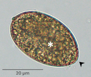
Egg morphology can be useful when gravid proglottids containing eggs are recovered, or when eggs are recovered via centrifugal fecal flotation (Figure 3). Taeniid eggs contain a developed larva with 6 hooklets (hexacanth embryo) and have a striated protective shell. Both features are detectable by light microscopy. In Mesocestoides species eggs, the outer shell is a thin membrane. Eggs of D caninum are released in characteristic egg packets (Figure 4). Diphyllobothriid eggs are distinguished by the presence of an operculum and an undeveloped larva and can be mistaken for trematode eggs.


Other Diagnostic Methods
Other diagnostic methods used to detect tapeworm infections in dogs and cats include antigen detection in feces and DNA amplification (copro-PCR).34,35 The use of the latter is of particular importance to differentiate Taenia eggs from Echinococcus eggs due to the zoonotic relevance of Echinococcus species. Morphology cannot differentiate these 2 genera, and their eggs are often generally referred to as taeniid eggs.
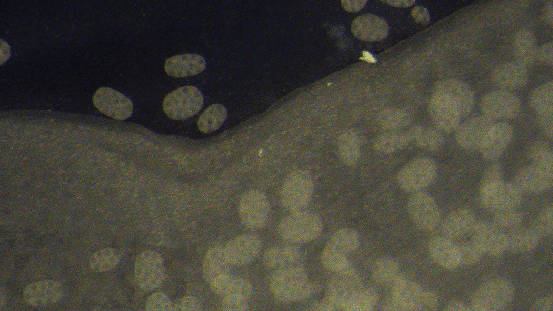
Figure 4. Dipylidium caninum gravid proglottid releasing egg packets
Prevention and Treatment Options
Because cestode infection involves 1 or 2 intermediate hosts, prevention requires limiting predation, eliminating intermediate hosts when possible, and providing anticestodal drugs according to exposure risks. It is important to note that praziquantel resistance has been reported in D caninum infection. Extralabel use of nitroscanate (50 mg/kg PO q24h in dogs) and nitazoxanide (100 mg/kg PO q24h in dogs and cats) has proved to be effective in such cases.36,37 For more information on treating cestode infections, see Resource 1.
Summary
Adult cestode infections in dogs and cats are usually asymptomatic and well tolerated; however, pet owners commonly observe tapeworm segments in pet resting areas or feces, causing discomfort and zoonotic concerns. Appropriate identification of these parasites is critical to disrupt their life cycle, provide effective treatment, and avoid reinfection. Learning how to identify the most common tapeworms in clinics improves diagnostic accuracy.
References
- Conboy G. Cestodes of dogs and cats in North America. Vet Clin North Am Small Anim Pract. 2009;39(6):1075-1090. doi:10.1016/j.cvsm.2009.06.005
- Králová-Hromadová I, Radačovská A, Čisovská Bazsalovicsová E, Kuchta R. Ups and downs of infections with the broad fish tapeworm Dibothriocephalus latus in Europe from 1900 to 2020: part I. Adv Parasitol. 2021;114:75-166. doi:10.1016/bs.apar.2021.08.008
- Scholz T, Kuchta R, Brabec J. Broad tapeworms (Diphyllobothriidae), parasites of wildlife and humans: recent progress and future challenges. Int J Parasitol Parasites Wildl. 2019;9:359-369. doi:10.1016/j.ijppaw.2019.02.001
- Sanders TL, Sobotyk C, Jimenez Castro PD, et al. Molecular characterization of Spirometra isolates across the USA. Parasitology. 2025;152(5):477-486. doi:10.1017/S003118202500054X
- Labuschagne M, Beugnet F, Rehbein S, Guillot J, Fourie J, Crafford D. Analysis of Dipylidium caninum tapeworms from dogs and cats, or their respective fleas – part 1. Molecular characterization of Dipylidium caninum: genetic analysis supporting two distinct species adapted to dogs and cats. Parasite. 2018;25:30. doi:10.1051/parasite/2018028
- Jesudoss Chelladurai JRJ, Abraham A, Quintana TA, Ritchie D, Smith V. Comparative genomic analysis and species delimitation: a case for two species in the zoonotic cestode Dipylidium caninum. Pathogens. 2023;12(5):675. doi:10.3390/pathogens12050675
- Beugnet F, Labushagne M, de Vos C, Crafford D, Fourie J. Analysis of Dipylidium caninum tapeworms from dogs and cats, or their respective fleas. Parasite. 2018;25(31):1-7. doi:10.1051/parasite/2018029
- Gates MC, Nolan TJ. Endoparasite prevalence and recurrence across different age groups of dogs and cats. Vet Parasitol. 2009;166(1-2):153-158. doi:10.1016/j.vetpar.2009.07.041
- Bowman DD, Hendrix CM, Lindsay DS, Barr SC. Dipylidium caninum. In: Bowman DD, Hendrix CM, Lindsay DS, Barr SC, eds. Feline Clinical Parasitology. Iowa State University Press; 2002:205-211.
- Wilcox RS, Bowman DD, Barr, SC, Euclid JM. Intestinal obstruction caused by Taenia taeniaeformis infection in a cat. JAAHA. 2009;45(2):93-96. doi:10.5326/0450093
- McGarry J, Collins M, Baross K. UK report of tapeworm. Vet Rec. 2020;186(15):498-499. doi:10.1136/vr.m1689
- Skirnisson K, Jouet D, Ferté H, Nielsen ÓK. Occurrence of Mesocestoides canislagopodis (Rudolphi, 1810) (Krabbe, 1865) in mammals and birds in Iceland and its molecular discrimination within the Mesocestoides species complex. Parasitol Res. 2016;115(7):2597-2607. doi:10.1007/s00436-016-5006-5
- Jesudoss Chelladurai JR, Brewer MT. Global prevalence of Mesocestoides infections in animals: a systematic review and meta-analysis. Vet Parasitol. 2021;298:109537. doi:10.1016/j.vetpar.2021.109537
- Wirtherle N, Wiemann A, Ottenjann M, et al. First case of canine peritoneal larval cestodosis caused by Mesocestoides lineatus in Germany. Parasitol Int. 2007;56(4):317-320. doi:10.1016/j.parint.2007.06.006
- Venco L, Kramer L, Pagliaro L, Genchi C. Ultrasonographic features of peritoneal cestodiasis caused by Mesocestoides sp. in a dog and cat. Vet Radiol Ultrasound. 2005;46(5):417-422. doi:10.1111/j.1740-8261.2005.00076.x
- Eleni C, Scaramozzino P, Busi M, Ingrosso S, D’Amelio S, De Liberato C. Proliferative peritoneal and pleural cestodiasis in a cat caused by metacestodes of Mesocestoides sp. Anatomohistopathological findings and genetic identification. Parasite. 2007;14(1):71-76. doi:10.1051/parasite/2007141071
- Boyce W, Shender L, Schultz L, et al. Survival analysis of dogs diagnosed with canine peritoneal larval cestodiasis (Mesocestoides spp.). Vet Parasitol. 2011;180(3-4):256-261. doi:10.1016/j.vetpar.2011.03.023
- Lanteri G, di Caro G, Capucchio M, et al. Mesocestoides and multivisceral tetrathyridiosis in a European cat. Veterinární Medicína. 2017;62(6):356-362. doi:10.17221/6/2017-vetmed
- Carta S, Corda A, Tamponi C, et al. Clinical forms of peritoneal larval cestodiasis by Mesocestoides spp. in dogs: diagnosis, treatment and long term follow-up. Parasitol Res. 2021;120(5):1727-1735. doi:10.1007/s00436-021-07107-w
- Alvarez Rojas CA, Romig T, Lightowlers MW. Echinococcus granulosus sensu lato genotypes infecting humans – review of current knowledge. Int J Parasitol. 2014;44(1):9-18. doi:10.1016/j.ijpara.2013.08.008
- Lymbery AJ, Jenkins EJ, Schurer JM, Thompson RCA. Echinococcus canadensis, E. borealis, and E. intermedius. What’s in a name? Trends Parasitol. 2015;31(1):23-29. doi:10.1016/j.pt.2014.11.003
- Chaâbane-Banaoues R, Oudni-M’rad M, Cabaret J, M’rad S, Mezhoud H, Babba H. Infection of dogs with Echinococcus granulosus: causes and consequences in an hyperendemic area. Parasit Vectors. 2015;8:231. doi:10.1186/s13071-015-0832-3
- Almulhim AM, John S. Echinococcus granulosus. StatPearls. Updated August 7, 2023. Accessed March 12, 2026. https://www.ncbi.nlm.nih.gov/books/NBK539751
- Evason MD, Peregrine AS, Jenkins EJ, et al. Emerging Echinococcus tapeworms: fecal PCR detection of Echinococcus multilocularis in 26 dogs from the United States and Canada (2022–2024). JAVMA. 2025;263(2):1-5. doi:10.2460/javma.24.07.0471
- Langs Rund L, Evason MD, Weese JS, Leutenegger CM. Echinococcus multilocularis in a cat: novel report of molecular detection (quantitative polymerase chain reaction) and management in a domestic cat from Canada. Am J Vet Res. 2025;86(7):ajvr.25.02.0072. doi:10.2460/ajvr.25.02.0072
- Umhang G, Forin-Wiart Ma, Hormaz V, et al. Echinococcus multilocularis detection in the intestines and feces of free-ranging domestic cats (Felis s. catus) and European wildcats (Felis s. silvestris) from northeastern France. Vet Parasitol. 2015;214(1-2):75-79. doi:10.1016/j.vetpar.2015.06.006
- Karamon J, Sroka J, Dąbrowska J, et al. First report of Echinococcus multilocularis in cats in Poland: a monitoring study in cats and dogs from a rural area and animal shelter in a highly endemic region. Parasit Vectors. 2019;12(1):313. doi:10.1186/s13071-019-3573-x
- Simoncini A, Massolo A. Multiscale ecological drivers of Echinococcus multilocularis spatial distribution in wild hosts: a systematic review. Food Waterborne Parasitol. 2023;34:e00216. doi:10.1016/j.fawpar.2023.e00216
- Kolapo TU, Hay A, Gesy KM, et al. Canine alveolar echinococcosis: an emerging and costly introduced problem in North America. Transbound Emerg Dis. 2023;2023:5224160. doi:10.1155/2023/5224160
- Zajac A, Fairman D, McGee E, et al. Alveolar echinococcosis in a dog in the eastern United States. J Vet Diagn Invest. 2020;32(5):742-746. doi:10.1177/1040638720943842
- Williams LA, Walzthoni N. Diagnosis, treatment, and outcome of four dogs with alveolar echinococcosis in the northwestern United States. JAVMA. 2023;261(7):1-6. doi:10.2460/javma.22.12.054
- Drake DA, Carreno A, Blagburn BL, et al. Proliferative sparganosis in a dog. JAVMA. 2008;233(11):1756-1760. doi:10.2460/javma.233.11.1756
- Tokiwa T, Fushimi M, Chou S, et al. Aberrant sparganosis in cat caused by Spirometra mansoni (Cestoda: Diphyllobothriidae): a case report. BMC Vet Res. 2024;20(1):148. doi:10.1186/s12917-024-03995-z
- Leutenegger CM, Lozoya CE, Tereski J, et al. Comparative study of a broad qPCR panel and centrifugal flotation for detection of gastrointestinal parasites in fecal samples from dogs and cats in the United States. Parasit Vectors. 2023;16(1):288. doi:10.1186/s13071-023-05904-z
- Little S, Braff J, Duncan K, et al. Diagnosis of canine intestinal parasites: Improved detection of Dipylidium caninum infection through coproantigen testing. Vet Parasitol. 2023;324:110073. doi:10.1016/j.vetpar.2023.110073
- Jesudoss Chelladurai J, Kifleyohannes T, Scott J, Brewer MT. Praziquantel resistance in the zoonotic cestode Dipylidium caninum. Am J Trop Med Hyg. 2018;99(5):1201-1205. doi:10.4269/ajtmh.18-0533
- Santhakumari Manoj RR. Current perspectives regarding Dipylidium caninum infection in companion animals. Todays Vet Pract. 2023;13(4):22-26.