
To take the CE quiz, click here.
This quiz is open until August 2028.
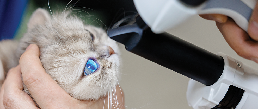
Corneal ulcers are among the most common ophthalmic diseases diagnosed in feline practice. Their treatment approach differs from that of corneal ulcers in dogs due to differences in etiology, management options, complications, and prognosis. Many corneal ulcers in cats, including both acute and chronic nonhealing superficial corneal ulcers, are directly or indirectly secondary to feline herpesvirus 1; identifying the specific etiology is essential for guiding appropriate treatment. This article focuses on the diagnosis of feline corneal ulcers based on eye examination findings and adjunctive testing, as well as the medical and surgical management of specific types of corneal ulcers.
Take-Home Points
- Feline herpesvirus 1 (FHV-1) is a common corneal ulcer etiology in cats and can be presumed unless another cause is identified.
- There is no reliable diagnostic test for FHV-1. Herpesvirus is often diagnosed based on clinical signs and response to therapy rather than laboratory testing.
- Superficial nonhealing corneal ulcers with loose epithelium can be secondary to FHV-1 in cats, requiring mechanical interventions (e.g., cotton-tipped applicator debridement, diamond burr debridement, superficial keratectomy) in conjunction with antiviral therapy to heal.
- Corneal sequestrum is a common complication in cats with nonhealing ulcers that confounds treatment. Prompt identification and treatment of ulcers are important to avoid this complication.
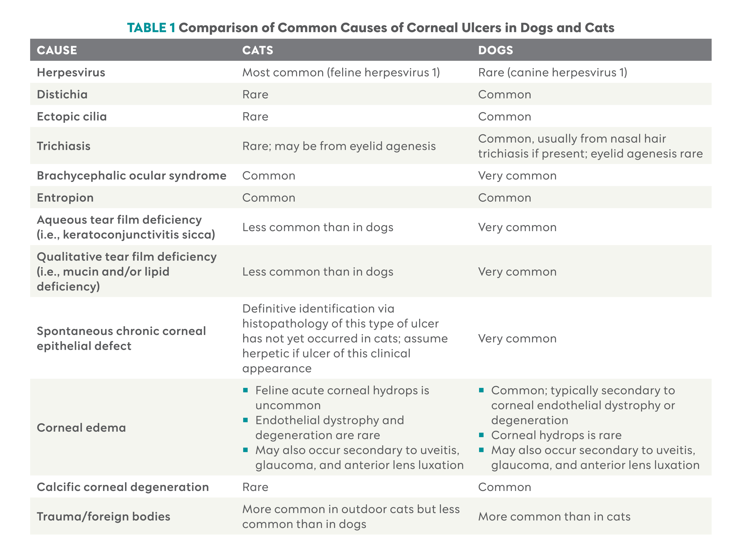
The cornea is an avascular, transparent structure. In dogs and cats, it is roughly 0.6 mm thick, consisting of 90% stromal collagen covered by a 6- to 10‑layer epithelium anteriorly and the endothelium–Descemet’s membrane complex posteriorly.1,2 Despite similar anatomy in the 2 species, the most common corneal diseases differ between dogs and cats, particularly in etiology and treatment of corneal ulceration (Table 1).
Etiologies
Epithelium is normally anchored to its basement membrane and anterior stroma and undergoes continuous renewal, with complete turnover estimated every 1 to 2 weeks. Corneal ulcers result from epithelial loss, exposing the underlying stroma. Etiologies of corneal ulcers can be divided into 3 categories:
- Mechanical epithelial removal: These cases include entropion, trichiasis, distichia, ectopic cilia, foreign bodies, and trauma. While common in dogs, these are rare in cats, except entropion, which is common in both species.3<.sup>
- Increased epithelial vulnerability: These patients have epithelial loss due to impaired corneal health (e.g., dry eye, brachycephalic ocular syndrome, facial nerve paralysis, buphthalmos, corneal edema). Brachycephalic ocular syndrome is common in both dogs and cats; otherwise, these causes are less common in cats.3
- Spontaneous epithelial loss: Epitheliotropic feline herpesvirus 1 (FHV-1) is the most common cause of corneal ulcers in cats. Spontaneous chronic corneal epithelial defects (SCCEDs), or indolent ulcers, are common in dogs but have not yet been definitively demonstrated to occur from the same pathophysiology in cats.
Secondary Infection
Epithelial loss compromises corneal defenses, which can allow opportunistic pathogens to invade and colonize the stroma at any time. The most common pathogens in cats are Staphylococcus species, followed by Streptococcus, Pseudomonas, Bacillus, or Pasteurella species.3-6 Proteolytic enzymes from bacteria and inflammatory cells cause stromal digestion, ulcer deepening, and widening. Descemetoceles occur when stromal loss exposes the Descemet’s membrane. Keratomalacia (melting ulcer) occurs when proteolytic enzymes induce rapid stromal liquefaction. Corneal perforation is a possibility once an ulcer is deep enough to compromise mechanical integrity, anecdotally considered a concern once the stromal loss begins to exceed 50% depth. Severe reflex uveitis (e.g., hypopyon, hyphema) can accompany infected ulcers.
Diagnosis
Ulcer diagnosis requires fluorescein staining to confirm epithelial loss, as well as identification of the underlying etiology and any secondary infection through a thorough ocular examination.
History and Signalment
Brachycephalic cats are at increased risk of ulcers.7 Maine coons, males, young adults, and middle‑aged to older cats are predisposed to entropion.8 A history of stress, immunosuppression, corticosteroid use, concurrent systemic disease, prior ulcers, conjunctivitis, or upper respiratory signs supports FHV‑1 involvement. A history of corticosteroid or cyclosporine therapy may be noted with feline acute corneal hydrops (also known as acute feline bullous keratopathy).9,10
Ocular Examination
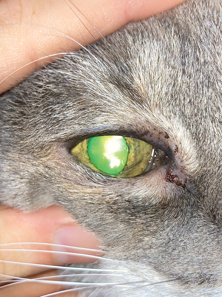
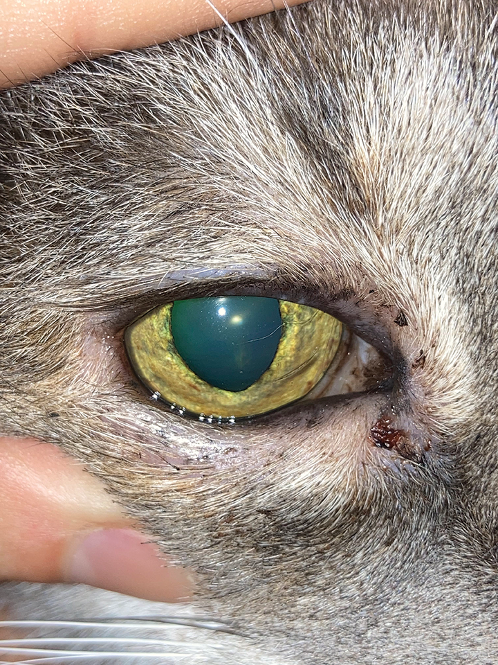
A complete ocular examination includes neuro-ophthalmic examination, conformation assessment (e.g., evaluating lower eyelid position to rule out entropion [Figure 1] and the severity of brachycephalic ocular features), and assessment of ocular structures (e.g., eyelids, cornea, intraocular structures) using magnification and a light source.
Screen for the following:
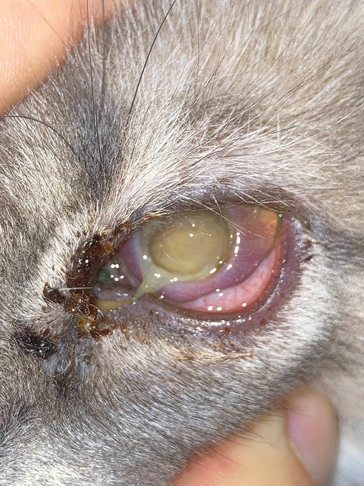
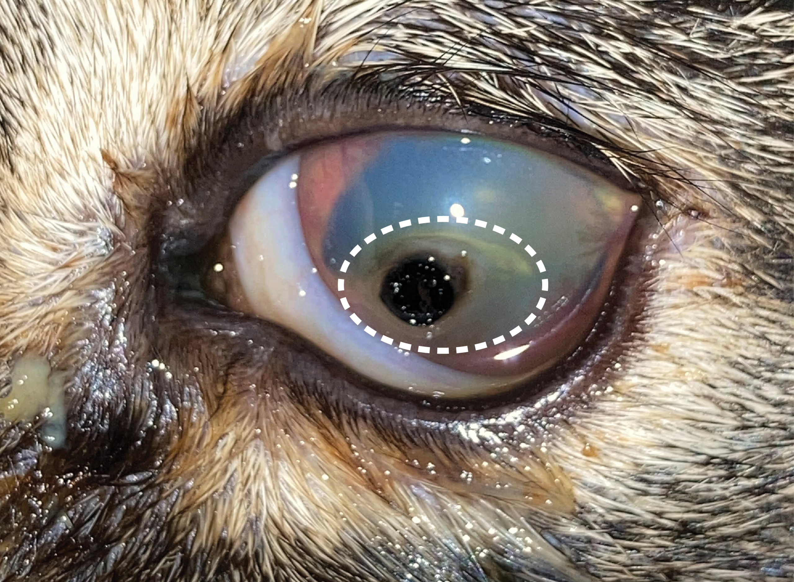
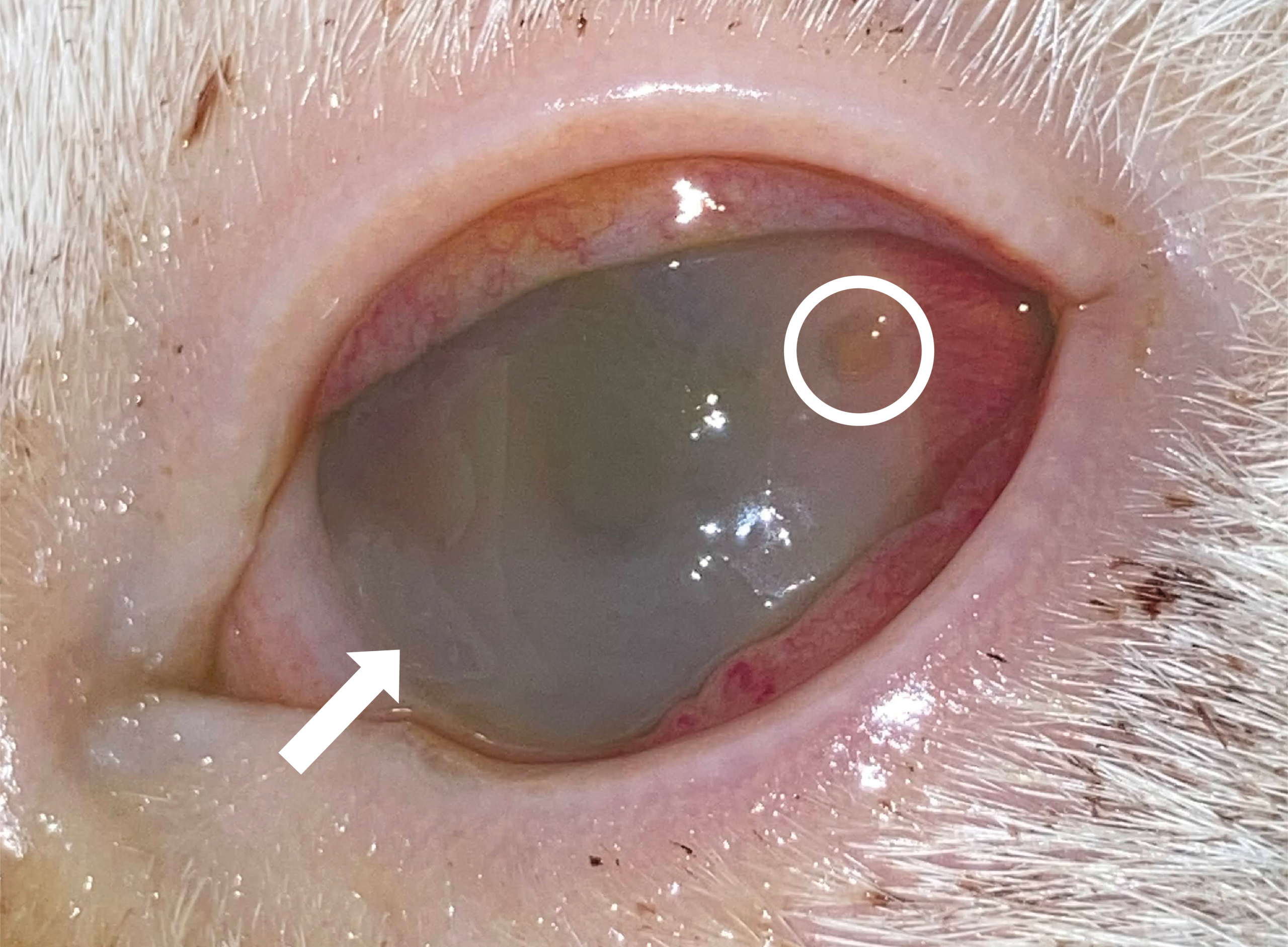
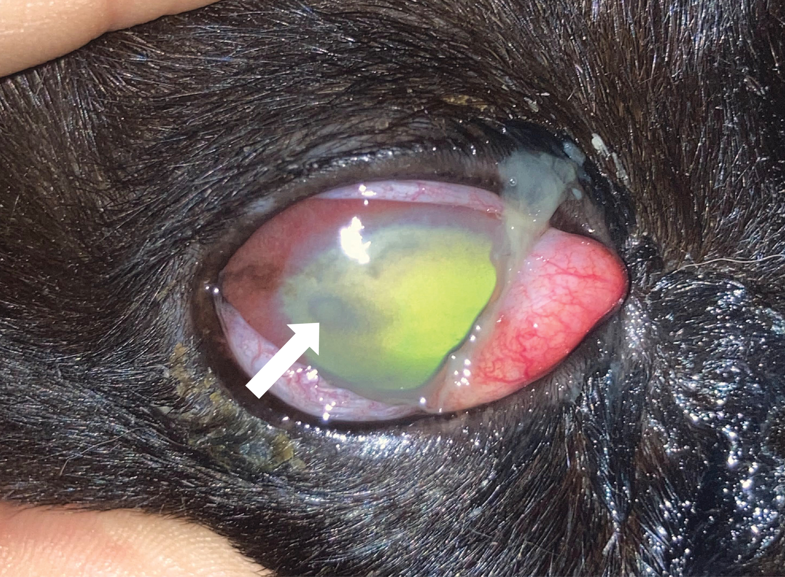
- Stromal loss (i.e., corneal flattening or depression [Figures 2–4]), yellow-white infiltrates (Figures 2, 4, and 5), keratomalacia (i.e., soft, sagging, gelatinous cornea [Figure 4]), or uveitis (e.g., flare, hypopyon, hyphema, iritis). Assume secondary infection if any of these changes are noted.
- A focal, raised area, made up of coagulated fibrin or iris, indicating a corneal perforation (Figure 6).
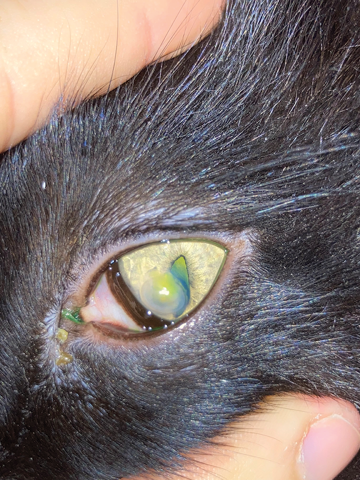
Figure 6. Corneal perforation. Note the irregular, protruding corneal surface with dyscoria from anterior iris prolapse, attempting to plug the defect.
- Loose epithelium, analogous to a wrinkled bedsheet or the peeling outer layer of onion skin, consistent with a nonhealing indolent ulcer (Figures 7 and 8).
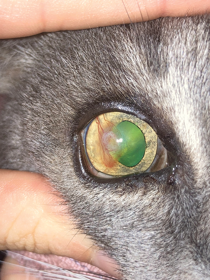
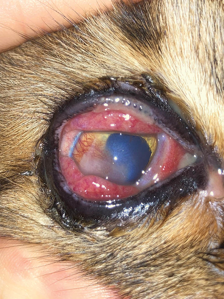
- Corneal neovascularization, which indicates chronicity, as it typically takes a few days to form, starting at the limbus and progressing further into the cornea with time.11 Dense, nonbranching, paintbrush-like vessels indicate deep disease (e.g., deep corneal ulcer, deep corneal infection [Figures 2, 4, and 5]) and thin, branching vessels indicate superficial disease (e.g., superficial ulcer [Figures 7–9]).
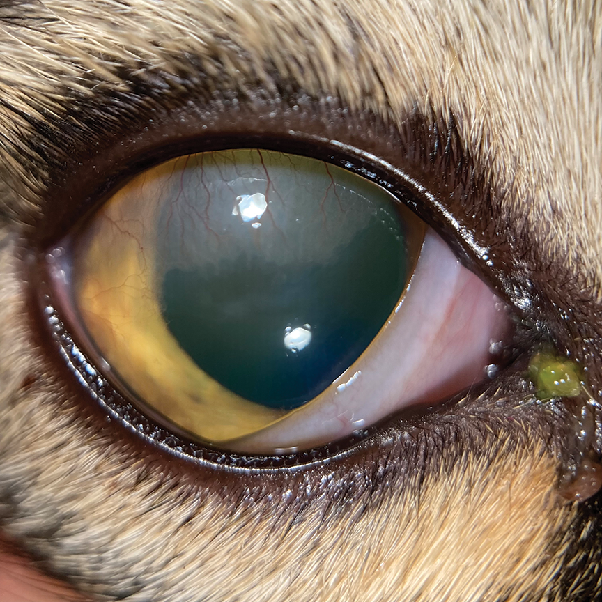
Figure 9. Chronic, superficial, herpetic ulcer with corneal opacification and branching superficial neovascularization. The corneal opacity and irregularity are secondary to loose epithelium.
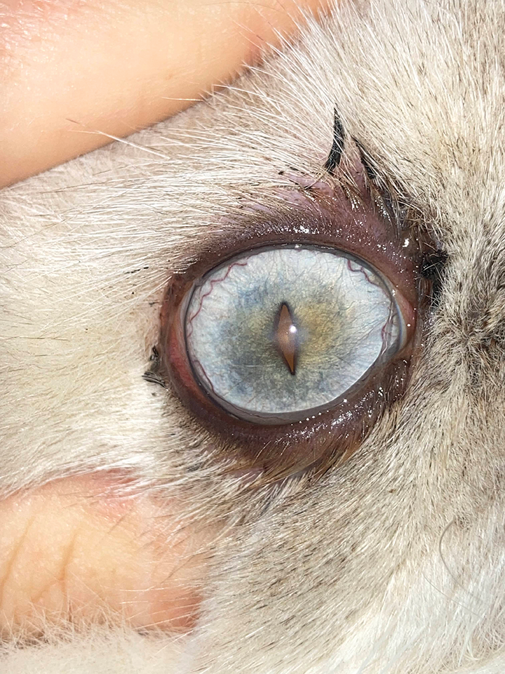
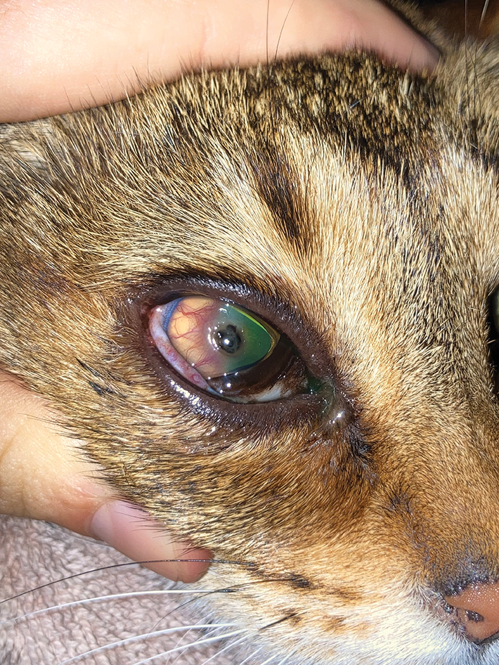
- Corneal sequestrum formation (i.e., a tea-stained or dark brown to black discoloration of the cornea [Figures 3, 10, and 11]).
- White-to-pink plaques, which are suggestive of eosinophilic keratitis.
- Blue-to-white cobblestoning, which indicates corneal edema. This finding can be seen in chronic superficial ulcers, where epithelial loss and neovascularization allow stromal hydration (Figure 8) and deep or infected ulcers. It is suspicious for feline acute corneal hydrops if accompanied by gross corneal swelling and forward corneal bulging.
Adjunctive Tests
Schirmer Tear Test
The Schirmer tear test (STT) measures aqueous tear production and is indicated for patients with chronic/recurrent ulcers or corneal neovascularization/opacification. Perform this test before applying any topical agents (e.g., fluorescein). Diagnosis of feline tear deficiency requires an STT < 9 mm/min on 2 separate occasions with clinical signs (Figure 12).12,13
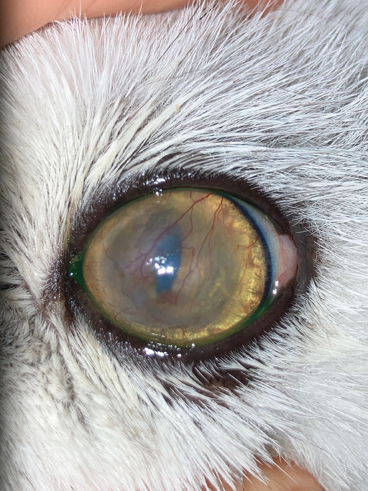
Figure 12. Aqueous tear deficiency. This patient had recurrent corneal ulcers, superficial neovascularization (thin, branching), and a lackluster tear film (note irregular flash artifact). Schirmer tear test results were < 9 mm/min on 2 separate occasions, confirming the diagnosis.
Fluorescein Staining
Fluorescein binds to exposed corneal stroma. Evaluate the cornea before fluorescein staining to avoid masking subtle infiltrates. Apply fluorescein stain to any eye with signs of ocular pain (e.g., blepharospasm, epiphora), redness, or opacity, then rinse excess and examine under cobalt blue light. Staining characteristics can suggest underlying etiologies, including:
- Mechanical epithelial removal. Stain location can be a key indicator to cause; axial staining is associated with brachycephalic ocular syndrome, ventrolateral with entropion, and ventromedial with third‑eyelid foreign bodies.7
- Herpetic ulcers. Typically geographic; dendritic patterns are rare but pathognomonic. Superficial ulcers with hazy, indistinct borders from stain migration underneath loose epithelium indicate FHV-1.
- Descemetoceles. No uptake at the center (exposed Descemet’s membrane); uptake occurs only in the surrounding stromal ring.
- Eosinophilic keratitis. Uptake on pink-to-white plaques does not indicate a true ulcer; uptake elsewhere indicates concurrent true ulceration, which is common with this disease.
- Feline acute corneal hydrops. Massive edema with a protruding cornea due to a spontaneously ruptured Descemet’s membrane that allows rapid and dramatic corneal hydration, which may be accompanied by corneal ulceration.10,14 Distinguish from melting ulcers, as treatment differs.
Seidel Test
The Seidel test is used to identify a leaking corneal perforation. Wet a fluorescein strip with a drop of sterile saline, then gently “paint” the suspicious area with the strip. This applies concentrated fluorescein stain. Do not rinse. Examine the area under cobalt blue light and magnification using a direct ophthalmoscope. Leaking aqueous humor creates a “waterfall” effect by diluting the stain.
Qualitative Tear Film Assessments
These tests identify lipid and/or mucin deficiencies, usually linked to FHV-1.15 Rose bengal (or lissamine green) stain identifies devitalized, mucin-deficient epithelium and early herpetic lesions in which some epithelial cell death is present, but the stroma is not exposed. The tear film break-up time test measures tear film stability, with values < 10 seconds indicating deficiency.12 Cochet–Bonnet esthesiometry can detect reduced corneal sensitivity, usually a metaherpetic change, and, while rarely performed, conjunctival biopsies can confirm mucin loss through reduced goblet cell density.15
Targeted Pathogen Identification
Paired corneal cytology and bacterial culture are indicated for potentially infected ulcers to identify bacteria and guide antibiotic therapy. If antibiotic susceptibility testing results are provided, interpret them cautiously; they are based on systemic concentrations and may not reflect in vivo efficacy of topical therapy, which can achieve ocular surface concentrations dramatically higher than serum levels.16,17 However, these high concentrations are rapidly diluted and washed away by the tear film, requiring repeated applications to maintain.
Herpesvirus Testing
Laboratory testing (e.g., serology, PCR) for FHV-1 is controversial and imprecise due to a variety of factors.18-20 For one, no diagnostic test can differentiate between wild-type and vaccine virus. In addition, false positives are possible because normal cats can shed the virus, and false negatives are also possible because diseased cats can shed the virus intermittently. Due to the challenges of test results, FHV-1 is typically diagnosed based on history, clinical signs, and response to therapy rather than laboratory testing.
Principles of Corneal Ulcer Treatment
Regardless of ulcer type, the fundamental management principles are consistent:
- Address the underlying cause.
- Provide appropriate topical antimicrobial therapy (prophylactic if infection is not present or intensive and targeted for an active infection).
- Control pain.
- Support healing.
Specific treatments vary with the ulcer’s etiology, depth, and chronicity and presence of infection.
Address the Underlying Cause
FHV-1 Ulcers
Any feline ulcer, including acute and chronic, superficial, nonhealing ulcers, can be presumed herpetic unless another cause is identified.
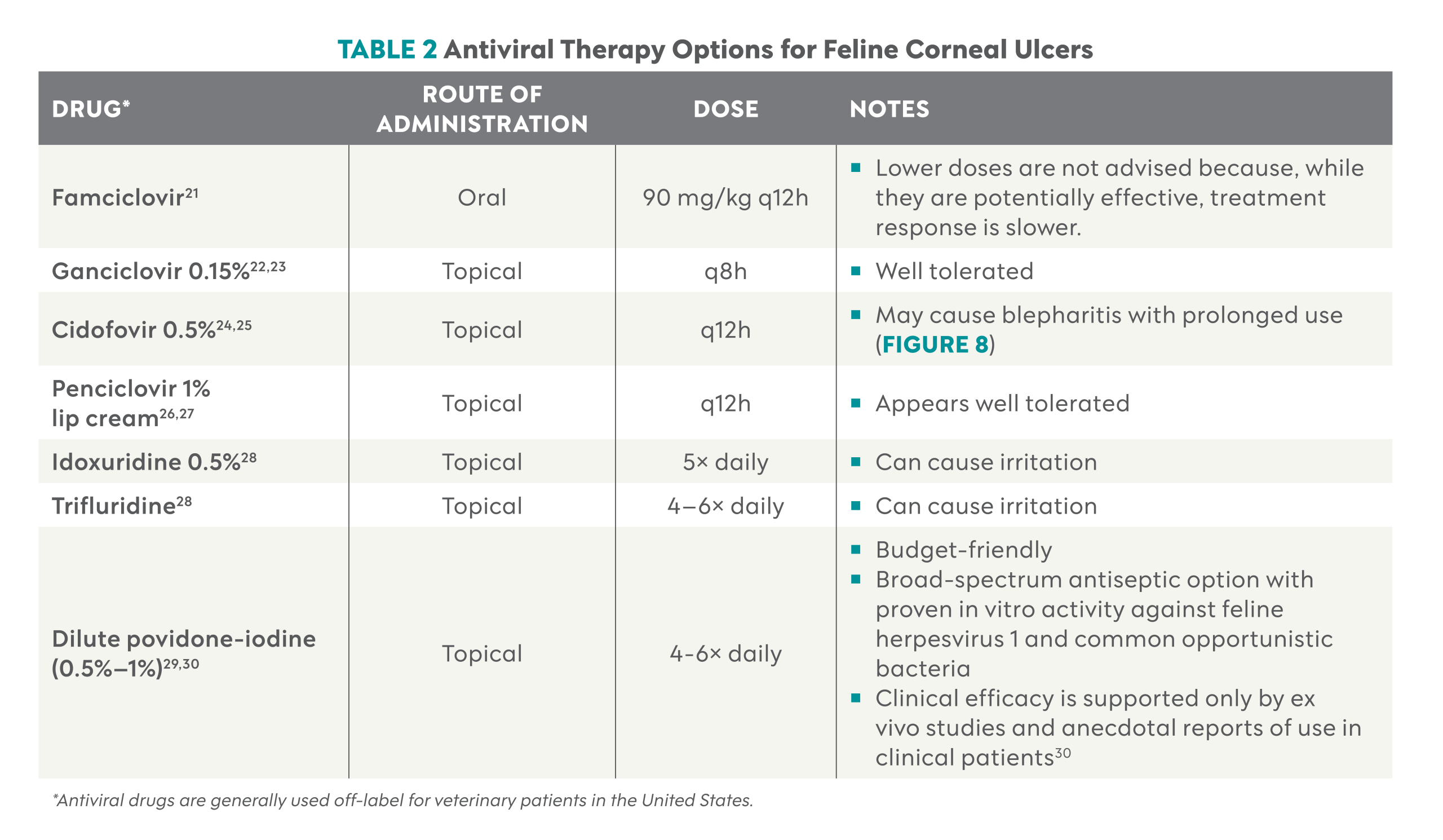
Prompt antiviral therapy speeds up healing and reduces complications (Table 2). Antivirals are virostatic, reducing viral load to allow the immune system to return the virus to dormancy. Continue antivirals 1 to 2 weeks beyond disease resolution. Lower dosing frequencies and the route (topical versus oral) best tolerated by the individual cat are preferred to minimize stress. Despite widespread use, lysine should not be relied upon as an effective antiviral to treat active herpetic disease, as evidence supporting its efficacy against FHV-1 is at best mixed.31-34
After topical anesthetic application, cotton‑tipped applicator (CTA) debridement of any loose, virus‑laden epithelium may promote healing of nonhealing superficial ulcers. It can also unmask ulcers when fluorescein staining appears negative (Figures 9 and 13) but epithelial irregularity and blepharospasm suggest an ulcer concealed by loose epithelium.
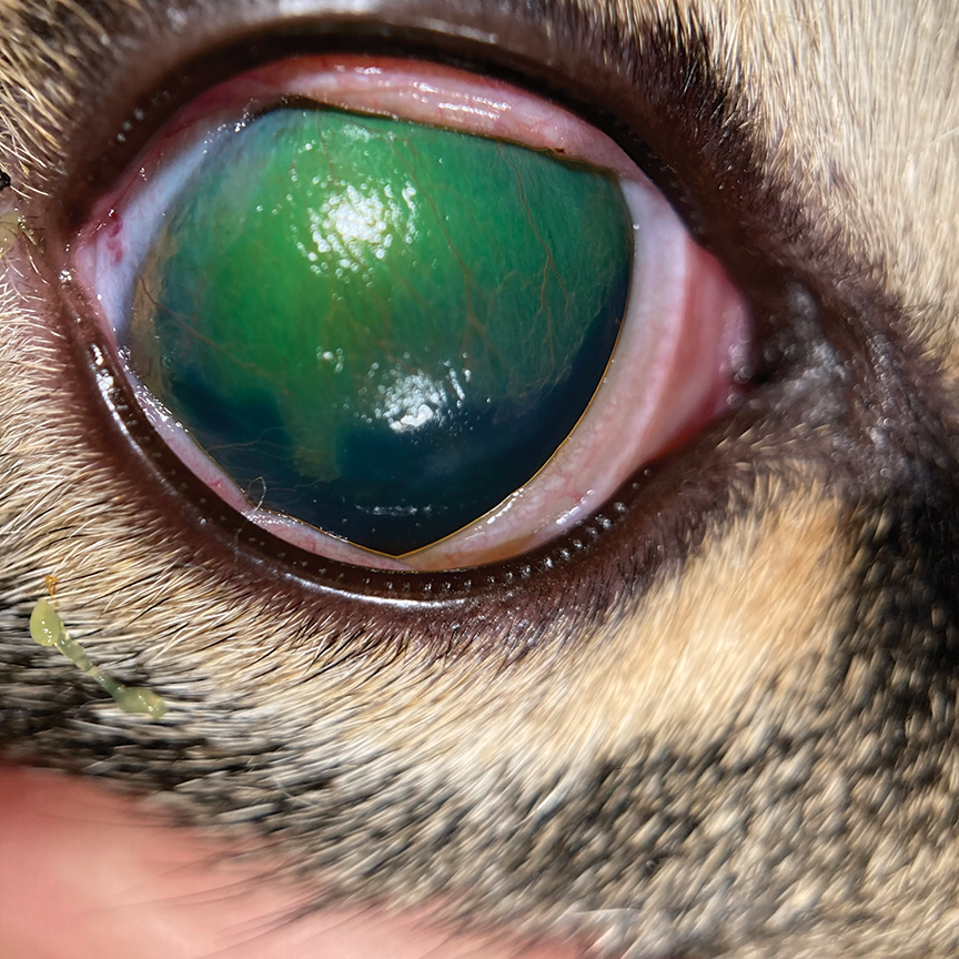
Figure 13. Same eye as Figure 9. After cotton-tipped applicator debridement, the true extent of the nonhealing ulcer is revealed.
Superficial, Nonhealing Ulcers With Loose Epithelium
Clinically, these resemble SCCEDs, also called indolent ulcers, seen in dogs; however, in cats, they are frequently, but not always, herpetic in origin.35-38 Despite the potential difference in pathogenesis between superficial, nonhealing ulcers with loose epithelium in dogs and cats, both are managed with interventions to stimulate healing:
- CTA debridement: A first-line intervention to remove loose epithelium that can be repeated every 2 to 3 weeks.36,38
- Diamond burr debridement: Noninvasive; indicated if CTA debridement fails (> 80% success rate at healing in 2 to 6 weeks).36,38
- Superficial keratectomy: Indicated for persistent cases or if there is concurrent sequestrum; requires referral (approximately 85% success rate at healing in 4 weeks).36,37
- Bandage contact lenses: Can be applied postprocedure and may help improve comfort and promote epithelialization.39,40
Due to the concern for these ulcers being caused by herpesvirus in cats and the difficulties in reliable diagnosis of herpesvirus via laboratory testing, concurrent antiviral therapy can be beneficial, but it may not be necessary for every cat.35-38
Importantly, these interventions are not appropriate in an infected ulcer, particularly if stromal loss is present. Additionally, unlike in dogs, where it is used to treat SCCEDs, grid keratotomy is contraindicated in cats due to the risk of corneal sequestrum formation.35
Entropion-Related Ulcers
Entropion is classified as primary/anatomic or secondary/spastic. With spastic entropion, the eyelid only rolls in when corneal pain is present. These are differentiated using topical anesthesia, which temporarily resolves spastic entropion. Unlike dogs, in which anatomic entropion is typically seen in juveniles, in cats, primary entropion occurs in both young adults and geriatric patients, often due to senile orbital fat loss and enophthalmos.
Surgical correction via the Hotz-Celsus procedure is recommended for anatomic entropion. In cats, concurrent lateral canthal closure can improve outcomes. In this procedure, a 2- to 3-mm long and 1- to 2-mm wide strip of both the upper and lower eyelids is resected at the lateral canthus, and the resected lid margins are sutured together in a single layer.41 This can help stabilize the eyelids to reduce the risk of entropion recurrence.41
Temporary entropion correction options include temporary tacking sutures for spastic cases and subdermal hyaluronic acid fillers as a nonsurgical alternative, performed under local anesthesia for mild to moderate senile entropion. Repeated injections of subdermal hyaluronic acid filler may be required as the product breaks down.42
Lubrication-Responsive Diseases
Topical lubrication with a sodium hyaluronate–containing gel (3× to 4× daily) is essential for cats with dry eye (aqueous, mucin, or lipid deficiencies) or brachycephalic ocular syndrome. In cats, FHV-1 is the primary cause of tear film deficiencies.13,15 Unlike dogs, cats respond poorly to lacrimostimulants and typically require tear replacement therapy alone.13 For brachycephalic cats, lubrication mitigates exposure-related irritation and inflammation. Medial canthoplasty to surgically reduce eyelid opening width and reduce corneal exposure is also useful.
In cats, avoid ointments and, ideally, preservative-containing products for long-term use (> 2 to 3 weeks) due to frequent hypersensitivity to lanolin, petrolatum, and preservatives.43 Cross-linked hyaluronic acid (i.e., sodium hyaluronate) gels are preferred for their superior ocular surface retention times (e.g., Ocunovis, Sentrx).44
Feline Acute Corneal Hydrops/Feline Acute Bullous Keratopathy
This condition is typically treated with a third eyelid flap to tamponade the cornea, which is left in place for 21 days. This procedure has an 85% success rate.10
Provide Antimicrobial Therapy
Antimicrobial therapy for corneal ulcers is either prophylactic for noninfected ulcers of any etiology or targeted and intensive if secondary infection is present.
Noninfected Ulcers
Prophylactic topical antibiotics with a spectrum that covers the most common secondary invading pathogens (e.g., tobramycin, chloramphenicol, erythromycin, chlortetracycline) are used 2 to 4 times daily. Fluoroquinolones and polymyxin-containing products should be avoided in noninfected ulcers to preserve antibiotic stewardship.45,46 Avoid using ointments for more than 2 to 3 weeks in cats.43
Although prophylactic antibiotic therapy is considered poor antibiotic stewardship, an exception is made in situations where the risk of infection is high and infection consequences would be catastrophic.47 Prophylactic therapy of corneal ulcers is considered to meet these criteria, and the current standard of care is to include topical antimicrobials in the treatment of noninfected ulcers. However, efforts can still be made to reduce antibiotic usage, such as considering antibiotic alternatives for prophylaxis of noninfected ulcers. This involves using products that have antibacterial activity against the most common pathogens that infect corneal ulcers but do not have the same risk of inducing antibiotic resistance.29,48 Such products can be applied 3 to 4 times daily and include the following:
- Cross-linked hyaluronic acid–containing gels. These gels are epitheliotropic and have bacteriostatic antimicrobial properties.49 To date, no studies have evaluated the efficacy of sodium hyaluronate–containing lubricants alone as prophylaxis for corneal ulcers in comparison with topical antibiotics.
- Ocular-specific antiseptic formulations. In vitro studies have demonstrated the efficacy of antiseptics against common pathogens involved in secondary bacterial infection of feline corneal ulcers, but follow-up in vivo studies are needed to demonstrate tolerability and efficacy.29 Available products contain 0.01% hypochlorous acid (e.g., Vetericyn Plus, Innovacyn) or 0.05% to 0.1% hexamidine diisethionate ± 0.0001% polyhexanide hydrochloride (e.g., Desomedine; Septostil, Trebifarma).29,50 Septostil, available in Europe, has been shown to be as effective as topical cefazolin at reducing the risk of infection after diamond burr debridement in dogs.50
Infected Ulcers
Dual antibiotic therapy is typically necessary to cover for common gram-positive bacteria such as Staphylococcus and Streptococcus species (e.g., chloramphenicol 0.5% to 1% solution, cefazolin 5% solution compounded from intravenous cefazolin vials) and gram-negative bacteria such as Pasteurella or Pseudomonas species (e.g., tobramycin or gentamicin 0.3% solution, ofloxacin or ciprofloxacin 0.3%). Initial dosing should be frequent (every 1 to 2 hours) for the first 24 to 72 hours to rapidly saturate the cornea and control infection, tapering once infection is controlled. Avoid antibiotic ointment formulations, since there is an increased risk of rupture from traumatic application and risk of granulomatous uveitis should the ointment enter the eye.
Systemic Antibiotics
Systemic antibiotics are rarely indicated for ulcers since they do not reach the infected site in the avascular cornea but may be justified in cases of perforation, deep ulcers at risk of rupture, or neovascularization reaching the infection site.
An exception is oral doxycycline, which is released in tears to reach the ocular surface; however, because the minimum inhibitory concentrations are lower than those of the most common corneal pathogens, its use is not recommended for antibiosis.51 It does have anticollagenolytic benefits, but equally effective nonantibiotic options are preferred (e.g., topical EDTA > 0.3%, serum/plasma, N-acetylcysteine > 0.5%) for antibiotic stewardship.52 N-Acetylcysteine is useful because it has biofilm-disrupting and broad-spectrum antimicrobial benefits.29,48 Dose topical anticollagenolytics every 1 to 2 hours until keratomalacia is controlled, then taper.
Adjunctive and Surgical Treatments
Noninvasive adjuncts to both medical and surgical management of infected or melting ulcers include corneal collagen cross-linking,53 which helps kill microorganisms rapidly and stabilize malacic cornea, and ultraviolet C light treatment, which kills microorganisms rapidly.54
Anecdotally, surgical stabilization may be indicated for ulcers exceeding 50% stromal depth to prevent rupture. A general rule is that if an ulcer has a concavity, it is at least 50% stromal depth. The surgical procedure typically involves keratectomy of necrotic and infected tissue. This excision of infected tissue facilitates rapid control of infection. The defect is then grafted for tectonic support. Graft options include autologous tissue (e.g., conjunctival pedicle grafts, corneoconjunctival transpositions) and biomaterials (e.g., amnion, porcine small intestinal mucosa). For deep, heavily infected ulcers, conjunctival pedicle grafts are ideal as they provide a direct blood supply, delivering endogenous antibiotic and anticollagenolytic factors. Notably, cats maintain superior corneal clarity postoperatively compared to dogs, especially if collagen-based or amniotic grafts are used rather than conjunctiva (Figure 14).55-57
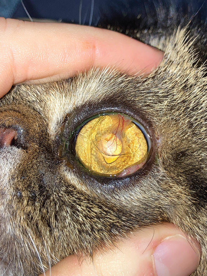
Figure 14. Excellent postoperative clarity in a cat 1 year after conjunctival pedicle graft for a deep, infected ulcer
Even with severe, deep, or perforated infected ulcers managed only medically, cats can have excellent outcomes.58 Compared to dogs with an equivalent ulcer, cats generally have a superior ability to heal. Hospitalization for intensive antibiotic therapy is recommended if owners cannot medicate at home and can be useful for close monitoring of a rapidly progressive ulcer. Enucleation is reserved for irreversibly blind eyes or to relieve pain in cases that do not improve after a week of intensive medical therapy.
Manage Pain
Effective analgesia is typically multimodal and maintained for the ulcer duration. There are no studies that specifically evaluate the efficacy of available feline pain medications for the treatment of corneal ulcer pain; therefore, this is an area where further research is needed. Instead, pain management targets corneal nerve pain and painful ciliary muscle spasm from reflex uveitis. Options include:
- Oral NSAIDs (e.g., low-dose meloxicam): Effective for corneal pain and associated reflex uveitis.
- Topical cycloplegics (e.g., 1% atropine): Controls ciliary spasm and stabilizes the blood–aqueous barrier. Dose every 24 to 48 hours for noninfected ulcers or every 8 to 12 hours initially for infected ulcers, tapering once mydriasis is achieved.
- Opioids (e.g., transmucosal buprenorphine): Useful for severe pain, after debridement, or following surgery.
- Adjuncts (e.g., gabapentin, pregabalin): May have benefits for corneal nerve pain, although analgesic efficacy and onset can be unpredictable; it may be best not to rely on as monotherapy.59 Anxiolytic effects may be useful during the treatment period.
Support Healing
While rigid plastic Elizabethan collars are standard for dogs, they should be used selectively in cats, primarily for fragile eyes or if self-trauma is observed. Since many feline ulcers are herpetic, the stress of a collar can exacerbate the condition and, in the authors’ experience, cats are less likely than dogs to self-traumatize. Avoid use of the following:
- Corticosteroids: Topical use is strictly contraindicated in any cat with a corneal ulcer. If fluorescein stain uptake is seen over a white plaque in a cat with eosinophilic keratitis, this is not true corneal ulceration and therefore topical steroids can be used to treat this immune-mediated disease; systemic use should also be avoided as it can contribute to FHV-1 reactivation or persistence.
- Topical NSAIDs: Generally avoid due to delayed healing, except when managing primary uveitis with secondary ulceration; for example, if a cat being treated with topical corticosteroids for uveitis has developed an ulcer from herpesvirus recrudescence. In such cases, switch to a topical NSAID to control the uveitis and stop the corticosteroid.
- Polymyxin-containing products: Linked to rare but potentially fatal anaphylaxis in cats.60
- Long-term (i.e., > 2 to 3 weeks) ointment use: Due to hypersensitivity concerns.43
- Ointment use in deep or infected ulcers: Due to challenges in administration and risk of granulomatous uveitis if the ointment enters the eye.
- Overtreatment: Excessive medications increase stress, promoting FHV-1 persistence. A minimalist protocol with only essential medications (e.g., antivirals, antimicrobials, pain management with or without lubrication when indicated) is preferred.
Prognosis
The prognosis for feline ulcers depends on the cause, duration, presence of infection, depth, and treatment choice.
Acute superficial ulcers typically heal within 5 to 7 days once the etiology is corrected. If there is concern for FHV-1, prompt antiviral therapy and CTA debridement of any loose epithelium are vital. Healing of FHV-1 ulcers can be more protracted. Recheck within 7 days to confirm resolution.
In cats with chronic superficial ulcers, loose epithelium requires intervention (e.g., CTA or diamond burr debridement, superficial keratectomy). Healing usually occurs within 2 weeks after the procedure; recalcitrant cases may take 4 to 6 weeks.36,38 Superficial keratectomy is the most successful surgical option; however, its disadvantages include the need for general anesthesia, higher costs, and the fact that the surgery involves the removal of approximately 20% of the corneal thickness.36,37 Therefore, it is not appropriate for every patient.36,37 Recheck every 10 to 14 days postprocedure.
FHV-1 is never cured; rather, it returns to dormancy. Therefore, stress-induced flare-ups and recurrent ulcers are common, especially in immunosuppressed, stressed, or systemically ill patients.36
In cats with bacterial infectious keratitis, the risk of rupture increases with ulcer depth. Surgical stabilization offers the most predictable outcomes, although many can heal with medical management alone.58 Monitor every 1 to 2 days until stable.
Common Complications
Ulcer Persistence
There is an impetus for rapid healing of feline ulcers. Delayed antiviral treatment for herpetic ulcers can lead to stalled healing and chronic ulcers that require intervention to obtain healing (e.g., diamond burr debridement, superficial keratectomy).
Fibrosis
Superficial ulcers typically heal without corneal fibrosis (scarring), whereas stromal ulcers result in disorganized collagen that leaves a gray-white opacity. While larger, deeper, or central scars impact vision more significantly, they can fade over time as collagen remodels. Notably, cats maintain corneal clarity after injury or surgery far better than dogs.55-57
Corneal Sequestrum
This is a complication unique to cats, common with ulcers that fail to heal rapidly.36,61 It is an area of necrotic, amber to black cornea triggered by chronic corneal irritation (Figures 3, 10, and 11). Controlled surgical excision of the corneal sequestrum and associated nonhealing ulcer is the preferred treatment, with additional grafting indicated if the excision bed is deep.61 Medical management is the alternative, waiting for the sequestrum to slough; however, this can take weeks to months and may result in persistent pain. Additionally, sequestra can deepen and enlarge, risking perforation after sloughing.61 Proactive intervention to promote rapid healing of feline ulcers is vital to prevent sequestrum formation. Unlike dogs, cats rarely develop pigmentary keratitis after corneal ulceration; they tend to form sequestra where a dog would develop pigmentary keratitis.
Corneoconjunctival Symblepharon
Corneoconjunctival symblepharon occurs when concurrent corneal and conjunctival ulceration causes apposed, inflamed surfaces to scar together, resulting in conjunctival bands or sheets covering the cornea (Figure 15). This complication typically follows primary FHV-1 infection in kittens.
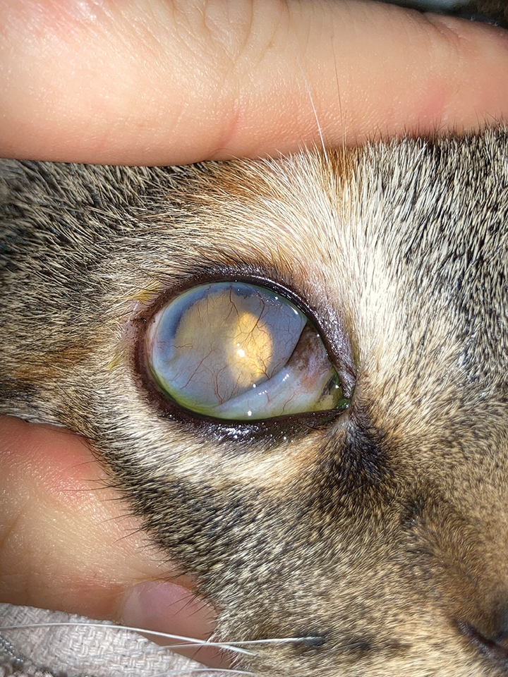
Figure 15. Severe corneal opacification in a cat secondary to corneoconjunctival symblepharon formation after severe primary herpesvirus ulcerative disease as a kitten. Note the veils of opaque and vascularized tissue covering the cornea.
Globe Perforation and Endophthalmitis
Globe perforation is a risk of infected ulcers. The deeper, wider, and more malacic the ulcer, the greater the risk. Surgical repair may restore integrity if a rupture occurs, and some ruptures can even heal medically if the defect is small enough to be stabilized by coagulated fibrin and/or iris (Figure 16).58 However, rupture can allow bacterial entry, potentially leading to endophthalmitis that often requires enucleation due to intractable inflammation and secondary glaucoma.
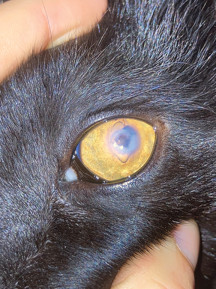
Figure 16. Corneal opacity secondary to corneal fibrosis and anterior synechiae (iris adhered to cornea) in a cat that was previously treated only medically for a perforated, infected corneal ulcer. The iris plugged the corneal defect at the time of perforation and became incorporated in the cornea during healing.
Summary
Appropriate corneal ulcer treatment in cats requires accurate diagnosis and prompt treatment of the underlying etiology (e.g., herpesvirus, dry eye, entropion), careful monitoring for complications (e.g., bacterial corneal infection, corneal sequestrum) and intervention should they develop, and variations in the medical and surgical treatment protocol depending on the type of ulcer (e.g., acute herpetic ulcer, superficial noninfected ulcer with loose epithelial edges, infected ulcer).
References
- Alario AF, Pirie CG. Central corneal thickness measurements in normal dogs: a comparison between ultrasound pachymetry and optical coherence tomography. Vet Ophthalmol. 2014;17(3):207-211. doi:10.1111/vop.12074
- Telle MR, Chen N, Shinsako D, et al. Relationship between corneal sensitivity, corneal thickness, corneal diameter, and intraocular pressure in normal cats and cats with congenital glaucoma. Vet Ophthalmol. 2019;22(1):4-12. doi:10.1111/vop.12558
- Verdenius CY, Broens EM, Slenter IJM, Djajadiningrat-Laanen SC. Corneal stromal ulcerations in a referral population of dogs and cats in the Netherlands (2012–2019): bacterial isolates and antibiotic resistance. Vet Ophthalmol. 2024;27(1):7-16. doi:10.1111/vop.13080
- Lin CT, Petersen-Jones SM. Antibiotic susceptibility of bacteria isolated from cats with ulcerative keratitis in Taiwan. J Small Anim Pract. 2008;49(2):80-83. doi:10.1111/j.1748-5827.2007.00437.x
- Goldreich JE, Franklin-Guild RJ, Ledbetter EC. Feline bacterial keratitis: clinical features, bacterial isolates, and in vitro antimicrobial susceptibility patterns. Vet Ophthalmol. 2020;23(1):90-96. doi:10.1111/vop.12693
- Suter A, Voelter K, Hartnack S, Spiess BM, Pot SA. Septic keratitis in dogs, cats, and horses in Switzerland: associated bacteria and antibiotic susceptibility. Vet Ophthalmol. 2018;21(1):66-75. doi:10.1111/vop.12480
- Frejlich M, Payen G. Prevalence and characteristics of feline ulcerative keratitis and corneal sequestra in a referral population and comparison between brachycephalic and nonbrachycephalic cats. Vet Ophthalmol. 2025;29(2):e70048. doi:10.1111/vop.70048
- Park J, Kang S, Lee E, Seo K. Prevalence of feline ophthalmic disorders in South Korea: a retrospective study (2009–2021). J Feline Med Surg. 2023;25(2):1098612X231151478. doi:10.1177/1098612X231151478
- Pierce KE, Wilkie DA, Gemensky-Metzler AJ, et al. An association between systemic cyclosporine administration and development of acute bullous keratopathy in cats. Vet Ophthalmol. 2016;19(S1):77-85. doi:10.1111/vop.12367
- Pederson SL, Pizzirani S, Andrew SE, Pate DO, Stine JM, Michau TM. Use of a nictitating membrane flap for treatment of feline acute corneal hydrops-21 eyes. Vet Ophthalmol. 2016;19(S1):61-68. doi:10.1111/vop.12353
- Anderson C, Zhou Q, Wang S. An alkali-burn injury model of corneal neovascularization in the mouse. J Vis Exp. 2014;(86):51159. doi:10.3791/51159
- Sebbag L, Kass PH, Maggs DJ. Reference values, intertest correlations, and test-retest repeatability of selected tear film tests in healthy cats. JAVMA. 2015;246(4):426-435. doi:10.2460/javma.246.4.426
- Uhl LK, Saito A, Iwashita H, Maggs DJ, Mochel JP, Sebbag L. Clinical features of cats with aqueous tear deficiency: a retrospective case series of 10 patients (17 eyes). J Feline Med Surg. 2019;21(10):944-950. doi:10.1177/1098612X18810867
- Schlesener BN, Scott EM, Vallone LV. An unusual case of feline acute corneal hydrops: atypical disease presentation and possible in vivo detection of Descemet’s membrane detachment in the cat’s unaffected eye. Vet Ophthalmol. 2018;21(4):426-431. doi:10.1111/vop.12527
- Cullen CL, Mjaa BL, Grahn BH. Ulcerative keratitis associated with qualitative tear film abnormalities in cats. Vet Ophthalmol. 1999;2(3):197-204. doi:10.1046/j.1463-5224.1999.00082.x
- Sigmund AB, Ward DA, Cox SK, Hendrix DVH. Tear film concentrations of topically applied 0.5% oxytetracycline ointment in normal canine eyes. Vet Ophthalmol. 2020;23(4):707-713. doi:10.1111/vop.12783
- Chitsaz S, Avizeh R, Najafzadeh Varzi H, Baniadam A. Comparison of serum oxytetracycline concentration after intravenous and intraosseous administration in dogs. Iranian Vet J. 2023;19(3):23-28. doi:10.22055/ivj.2023.365433.2511
- Maggs DJ, Clarke HE. Relative sensitivity of polymerase chain reaction assays used for detection of feline herpesvirus type 1 DNA in clinical samples and commercial vaccines. Am J Vet Res. 2005;66(9):1550-1555. doi:10.2460/ajvr.2005.66.1550
- Sjödahl-Essén T, Tidholm A, Thorén P, et al. Evaluation of different sampling methods and results of real-time PCR for detection of feline herpes virus-1, Chlamydophila felis and Mycoplasma felis in cats. Vet Ophthalmol. 2008;11(6):375-380. doi:10.1111/j.1463-5224.2008.00661.x
- Rampazzo A, Appino S, Pregel P, Tarducci A, Zini E, Biolatti B. Prevalence of Chlamydophila felis and feline herpesvirus 1 in cats with conjunctivitis in northern Italy. J Vet Intern Med. 2003;17(6):799-807. doi:10.1111/j.1939-1676.2003.tb02517.x
- Thomasy SM, Shull O, Outerbridge CA, et al. Oral administration of famciclovir for treatment of spontaneous ocular, respiratory, or dermatologic disease attributed to feline herpesvirus type 1: 59 cases (2006–2013). JAVMA. 2016;249(5):526-538. doi:10.2460/javma.249.5.526
- Lewin AC, Liu CC, Alling C, Camacho-Luna P, Miessler B, Carter RT. In vitro efficacy of ganciclovir against feline herpesvirus type 1 and assessment of ocular tolerability in healthy cats. J Feline Med Surg. 2021;23(4):400-404. doi:10.1177/1098612X20944363
- Ledbetter EC, Badanes ZI, Chan RX, et al. Comparative efficacy of topical ophthalmic ganciclovir and oral famciclovir in cats with experimental ocular feline herpesvirus-1 epithelial infection. J Ocul Pharmacol Ther. 2022;38(5):339-347. doi:10.1089/jop.2022.0001
- Ledbetter EC, Morgan AJ. Frequency and characterization of local ocular toxicity in cats treated with topical ophthalmic cidofovir for presumptive feline herpesvirus-1 infection. Vet Ophthalmol. 2026;29(1):e70046. doi:10.1111/vop.70046
- Fontenelle JP, Powell CC, Veir JK, Radecki SV, Lappin MR. Effect of topical ophthalmic application of cidofovir on experimentally induced primary ocular feline herpesvirus-1 infection in cats. Am J Vet Res. 2008;69(2):289-293. doi:10.2460/ajvr.69.2.289
- Ledbetter EC, Ramos-Bartolomei AM, Brito FLC, et al. Evaluation of topical ophthalmic application of penciclovir cream in cats with experimental ocular feline herpesvirus-1 infection. Am J Vet Res. 2026;87(3):ajvr.25.08.0304. doi:10.2460/ajvr.25.08.0304
- Pe’er O, Thomasy SM, Feuer E, Knych HK, Ofri R, Sebbag L. Ocular tolerance and tear film pharmacokinetics of 1% penciclovir cream in cats. Res Vet Sci. 2025;196:105909. doi:10.1016/j.rvsc.2025.105909
- Stiles J. Treatment of cats with ocular disease attributable to herpesvirus infection: 17 cases (1983-1993). JAVMA.
1995;207(5):599-603. - Wolff HT, Piroth AC, Oltmanns H, et al. Commercially available antiseptics show high in vitro efficacy against pathogens most commonly associated with canine and feline infectious keratitis. Front Vet Sci. 2025;12:1552230. doi:10.3389/fvets.2025.1552230
- Pennington MR, Capriotti JA, Van de Walle GR. In vitro efficacy of povidone iodine and hydroxyethyl cellulose, alone and in combination, against common feline ocular pathogens. Vet J. 2018;241:38-41. doi:10.1016/j.tvjl.2018.09.011
- Maggs DJ, Collins BK, Thorne JG, Naisisse MP. Effects of L-lysine and L-arginine on in vitro replication of feline herpesvirus type-1. Am J Vet Res. 2000;61(12):1474-1478. doi:10.2460/ajvr.2000.61.1474
- Stiles J, Townsend WM, Rogers QR, Krohne SG. Effect of oral administration of L-lysine on conjunctivitis caused by feline herpesvirus in cats. Am J Vet Res. 2002;63(1):99-103. doi:10.2460/AJVR.2002.63.99
- Cave NJ, Dennis K, Gopakumar G, Dunowska M. Effects of physiologic concentrations of l-lysine on in vitro replication of feline herpesvirus 1. Am J Vet Res. 2014;75(6):572-580. doi:10.2460/ajvr.75.6.572
- Drazenovich TL, Fascetti AJ, Westermeyer HD, et al. Effects of dietary lysine supplementation on upper respiratory and ocular disease and detection of infectious organisms in cats within an animal shelter. Am J Vet Res. 2009;70(11):1391-1400. doi:10.2460/ajvr.70.11.1391
- La Croix NC, van der Woerdt A, Olivero DK. Nonhealing corneal ulcers in cats: 29 cases (1991-1999). JAVMA. 2001;218(5):733-735. doi:10.2460/javma.2001.218.733
- Rujirekasuwan N, Sattasathuchana P, Sritrakoon N, Thengchaisri N. Recurrent vs. nonrecurrent superficial non-healing corneal ulcers in cats: a multifactorial retrospective analysis. Animals (Basel). 2025;15(14):2104. doi:10.3390/ani15142104
- Jégou JP, Tromeur F. Superficial keratectomy for chronic corneal ulcers refractory to medical treatment in 36 cats. Vet Ophthalmol. 2015;18(4):335-340. doi:10.1111/vop.12153
- Anastassiadis Z, Bayley KD, Read RA. Corneal diamond burr debridement for superficial non-healing corneal ulcers in cats. Vet Ophthalmol. 2022;25(6):476-482. doi:10.1111/vop.13026
- Bossuyt SM. The use of therapeutic soft contact bandage lenses in the dog and the cat: a series of 41 cases. Vlaams Diergeneeskd Tijdschr. 2016;85(6):343-348. https://doi.org/10.21825/vdt.v85i6.16308
- Dees DD, Fritz KJ, Wagner L, Paglia D, Knollinger AM, Madsen R. Effect of bandage contact lens wear and postoperative medical therapies on corneal healing rate after diamond burr debridement in dogs. Vet Ophthalmol. 2017;20(5):382-389. doi:10.1111/vop.12412
- White JS, Grundon RA, Hardman C, O’Reilly A, Stanley RG. Surgical management and outcome of lower eyelid entropion in 124 cats. Vet Ophthalmol. 2012;15(4):231-235. doi:10.1111/j.1463-5224.2011.00974.x
- McDonald JE, Knollinger AM. The use of hyaluronic acid subdermal filler for entropion in canines and felines: 40 cases. Vet Ophthalmol. 2019;22(2):105-115. doi:10.1111/vop.12566
- Eördögh R, Schwendenwein I, Tichy A, Loncaric I, Nell B. Clinical effect of four different ointment bases on healthy cat eyes. Vet Ophthalmol. 2016;19(S1):4-12. doi:10.1111/vop.12279
- Grego AL, Fankhauser AD, Behan EK, et al. Comparative fluorophotometric evaluation of the ocular surface retention time of cross-linked and linear hyaluronic acid ocular eye drops on healthy dogs. Vet Res Commun. 2024;48(6):4191-4199. doi:10.1007/s11259-024-10559-x
- AWaRe classification of antibiotics for evaluation and monitoring of use, 2023. World Health Organization. July 26, 2023. Accessed June 30, 2026. https://www.who.int/publications/i/item/WHO-MHP-HPS-EML-2023.04
- Schmerold I, Geijlswijk Iv, Gehring R. European regulations on the use of antibiotics in veterinary medicine. Eur J Pharm Sci. 2023;189:106473. doi:10.1016/j.ejps.2023.106473
- Frey E, Costin M, Granick J, Kornya M, Weese JS. 2022 AAFP/AAHA antimicrobial stewardship guidelines. JAAHA. 2022;58(4):1-5. doi:10.5326/1547-3317-58.4.1
- Walter H, Verspohl J, Meißner J, Oltmanns H, Geks AK, Busse C. In vitro antimicrobial activity of N-acetylcysteine against pathogens most commonly associated with infectious keratitis in dogs and cats. Antibiotics (Basel). 2023;12(3):559. doi:10.3390/antibiotics12030559
- Williams DL, Wirostko BM, Gum G, Mann BK. Topical cross-linked HA-based hydrogel accelerates closure of corneal epithelial defects and repair of stromal ulceration in companion animals. Invest Ophthalmol Vis Sci. 2017;58(11):4616-4622. doi:10.1167/iovs.16-20848
- Gimenez E, Crasta M. Comparision between the use of topical antibiotic and Septostil in spontaneous chronic corneal epithelial defects in dogs. Vet Ophthalmol. 2026;29(2):e70114. doi:10.1111/vop.70114
- Sebbag L, Showman L, McDowell EM, Perera A, Mochel JP. Impact of flow rate, collection devices, and extraction methods on tear concentrations following oral administration of doxycycline in dogs and cats. J Ocul Pharmacol Ther. 2018;34(6):452-459. doi:10.1089/jop.2018.0008
- Kimmitt BA, Moore GE, Stiles J. Comparison of the efficacy of various concentrations and combinations of serum, ethylenediaminetetraacetic acid, tetracycline, doxycycline, minocycline, and N-acetylcysteine for inhibition of collagenase activity in an in vitro corneal degradation model. Am J Vet Res. 2018;79(5):555-561. doi:10.2460/ajvr.79.5.555
- Crasta M, Gimenez E, Ostan PC, Arteaga K. Photoactivated chromophore for keratitis—corneal cross-linking in dogs and cats: a retrospective study in Italy. Vet Ophthalmol. 2025;28(2):413-424. doi:10.1111/vop.13284
- Turicea B, Sahoo DK, Allbaugh RA, Stinman CC, Kubai MA. Novel treatment of infectious keratitis in canine corneas using ultraviolet C (UV-C) light. Vet Ophthalmol. 2025;28(4):699-713. doi:10.1111/vop.13265
- Chow DWY, Westermeyer HD. Retrospective evaluation of corneal reconstruction using ACell Vet(™) alone in dogs and cats: 82 cases. Vet Ophthalmol. 2016;19(5):357-366. doi:10.1111/vop.12294
- Maini S, Hurley-Bennett K, Dawson C. Case series describing the use of low-temperature vacuum-dehydrated amnion (Omnigen) for the treatment of corneal ulcers in cats and dogs: 46 cases (2016-2017). Top Companion Anim Med. 2020;41:100474. doi:10.1016/j.tcam.2020.100474
- Goulle F. Use of porcine small intestinal submucosa for corneal reconstruction in dogs and cats: 106 cases. J Small Anim Pract. 2012;53(1):34-43. doi:10.1111/j.1748-5827.2011.01149.x
- Bustamante MGMd, Good KL, Leonard BC, et al. Medical management of deep ulcerative keratitis in cats: 13 cases. J Feline Med Surg. 2018;21(4):387-393. doi:10.1177/1098612X18770514
- Ciccarelli S, Perrone C, Forte L, Di Bello A, Franchini D. Efficacy of gabapentin as pain management of T2–T4 feline facial squamous cell carcinoma treated with electrochemotherapy. Sci Rep. 2025;15(1):27874. doi:10.1038/s41598-025-13767-w
- Hume-Smith KM, Groth AD, Rishniw M, Walter-Grimm LA, Plunkett SJ, Maggs DJ. Anaphylactic events observed within 4 h of ocular application of an antibiotic-containing ophthalmic preparation: 61 cats (1993–2010). J Feline Med Surg. 2017;13(10):744-751. doi:10.1016/j.jfms.2011.06.007
- Gómez AP, Mazzucchelli S, Smith K, de Lacerda RP. Long-term treatment outcomes and risk factors for recurrence in feline corneal sequestrum: 72 cases (2009–2017). Vet Rec. 2023;193(3):e2783. doi:10.1002/vetr.2783
CE Quiz
This article has been submitted for RACE approval for 1 hour of continuing education credit and will be opened for enrollment upon approval. To receive credit, take the test at vetfolio.com. Free registration is required. Questions and answers online may differ from those below. Tests are valid for 2 years from the date of approval.
1. What is the most likely etiology of a corneal ulcer in a cat if no other abnormalities are found on ocular examination?
a. Trauma
b. Bacterial infection
c. Feline herpesvirus 1 (FHV-1)
d. Tear deficiency
2. Which of the following findings is most indicative of a chronic corneal process?
a. Blepharospasm
b. Corneal neovascularization
c. Corneal edema
d. Conjunctival hyperemia
3. Which of the following is the best initial antibiotic treatment for a noninfected superficial corneal ulcer in a cat?
a. Topical fluoroquinolone every hour
b. Topical erythromycin ointment 2 to 4 times daily
c. Systemic antibiotic
d. Topical fluoroquinolone 2 to 4 times daily
4. A cat with suspected FHV-1 corneal ulceration is being treated. How long should antiviral therapy generally be continued after the ulcer has healed?
a. 1 to 2 weeks beyond clinical resolution
b. Until fluorescein stain is negative
c. Until clinical signs improve
d. Indefinitely
5. Which of the following is contraindicated in cats with superficial, nonhealing ulcers with loose epithelium?
a. Cotton-tipped applicator debridement
b. Diamond burr debridement
c. Superficial keratectomy
d. Grid keratotomy