
Fecal examination for parasite detection is a common laboratory procedure in veterinary practice as it is cost-effective and minimally invasive. These tests yield valuable information about patient wellness and the success of anthelmintic treatment. Several techniques, including fecal flotation, direct smear, and sedimentation, can be easily performed in-clinic; the results must be interpreted by trained and experienced personnel. Over the past decade, new fecal diagnostic tests for veterinary patients have become available, each with advantages and limitations. This article describes and compares fecal examination techniques currently offered by diagnostic laboratories to help clinicians choose the appropriate test based on each patient’s clinical situation.
Take-Home Points
- Regular fecal examination assesses efficacy of parasite prevention products and compliance of owners and allows detection of parasites not susceptible to monthly dewormers.
- Tests should be selected based on case differentials as well as test sensitivity and specificity. In general, select the procedure that gives the best chance of observing the parasite of interest if it is present.
- With the rising concern for anthelmintic resistance, quantitative assays are likely to be used more often in small animal medicine.
Endoparasites remain common in dogs and cats regardless of age, sex, breed, or history of preventive care, and fecal examination remains a primary diagnostic tool for both well and sick animals. According to recent surveys, hookworms (particularly Ancylostoma caninum) remain among the most common gastrointestinal worms of dogs, and Giardia duodenalis remains a prevalent problem in both dogs and cats.1,2 One study found at least 1 parasite in 20.7% of tested dogs attending parks, and more than 11% of adult dogs have parasites.1 Parasites are identified in 1 of 5 cats under 2 years of age, with more than 12% of cats under 5 years of age being positive for a parasite.2 Regular deworming helps control these infections, but no single product has efficacy against all parasites of pets, and anthelmintic resistance is a growing concern. For this reason, regular fecal testing—regardless of preventive use—should be performed to ensure detection of parasites that are not covered by monthly dewormers or that may be resistant to treatment.
Historically, samples were primarily processed in-clinic using passive or centrifugal fecal flotation methods, which allowed rapid delivery of results to clients; however, the sensitivity and specificity of in-clinic fecal flotation exams vary greatly according to microscopist training and type of assay (passive versus centrifugal). Veterinary teams with proper training in classic parasitology techniques can still perform selected parasitology diagnostic tests in-house.
In recent years, reference laboratories at universities or corporate facilities have risen in number and now provide further choices that can be used as primary tests or as adjunctive tools for specific scenarios (e.g., species identification, confirmation of anthelmintic resistance). Results can vary between laboratories, and if the results do not align with the clinical suspicion, consultation with a veterinary parasitologist from a veterinary college or diagnostic laboratory is recommended.
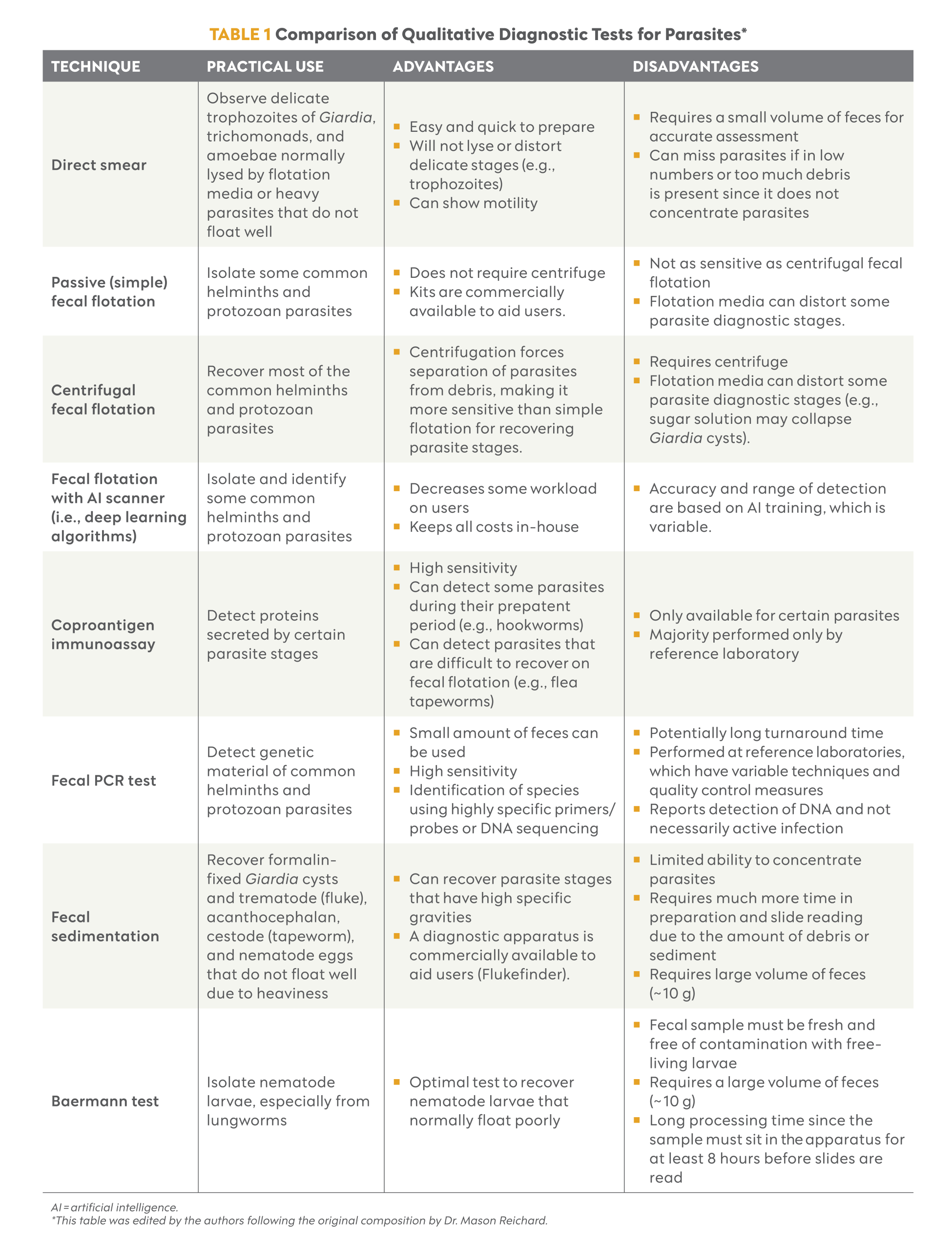
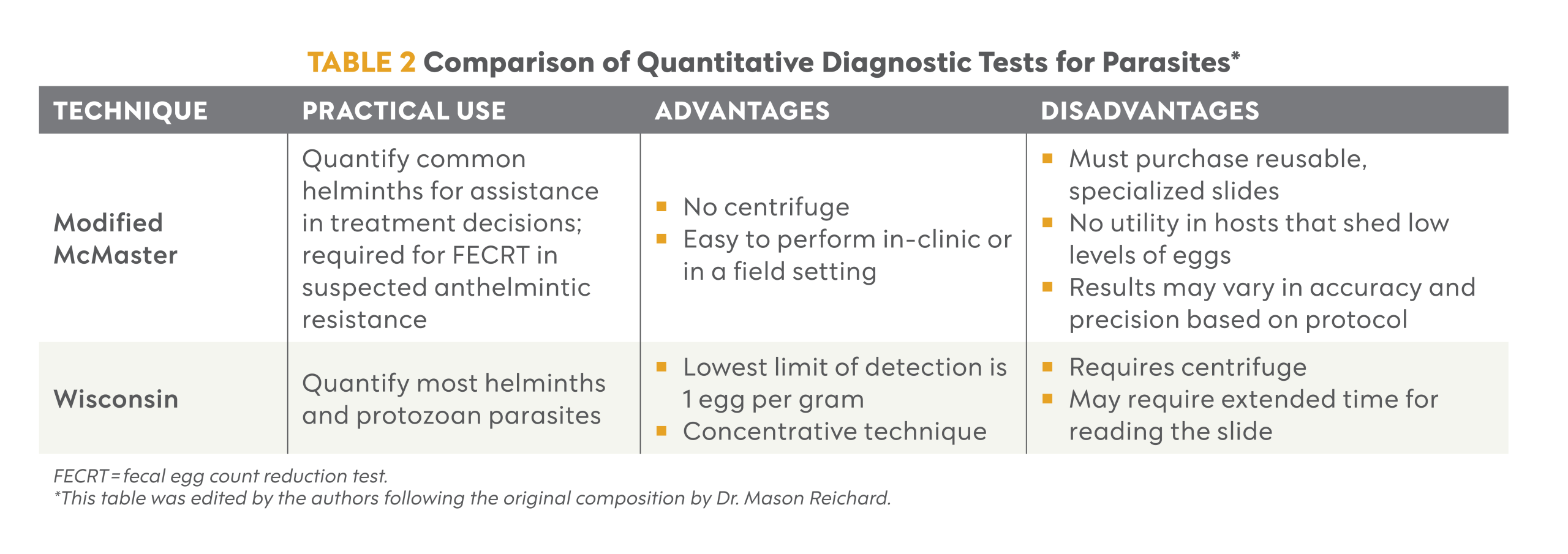
A single diagnostic laboratory may offer numerous tests for parasite detection in patient feces. For most cases, a qualitative test (Table 1) is adequate for determining the presence of a parasite in a small animal fecal sample. However, occasionally a quantitative exam (Table 2) is necessary when assessing treatment efficacy using a fecal egg count reduction test (FECRT).
Qualitative Fecal Examination Techniques
The advantages and limitations of currently available qualitative tests are briefly described below.
Direct Smear
Direct smears are performed using a small amount of feces with a drop of saline solution on a glass slide. Use of a small amount of feces is critical, as a thick layer will complicate correct assessment. The samples are examined under the microscope after applying a coverslip.
Direct smear is appropriate for the visualization of delicate trophozoites from Giardia, trichomonads, and amoebae.3,4 However, direct smears can be poorly sensitive, and the lack of recovered parasites should not discourage pursuing additional tests.
Fecal Flotation
Flotation techniques are used to visualize parasite eggs after mixing the fecal sample with a flotation solution. Fecal flotation can be performed by simply letting the mixture sit until the eggs float (passive flotation) or centrifuging the mixture (centrifugal flotation). Centrifugal flotation consistently recovers more eggs than passive flotation.5 Recently, a deep learning algorithm has been applied for the diagnosis of parasites in small animal pets.6-8
Most parasite eggs have a specific gravity (SG) that ranges between 1.05 and 1.23; therefore, the SG of the flotation solution must be higher than that of the target parasite egg.9 Common flotation solutions include zinc sulfate (SG 1.18 to 1.2), Sheather’s sugar (SG 1.2 to 1.27), sodium nitrate (SG 1.18 to 1.2), saturated salt (SG 1.18 to 1.2), and magnesium sulfate (SG 1.2).5 Zinc sulfate is preferred to visualize Giardia cysts without distorting them; however, an SG of 1.18 will recover fewer helminth eggs, including nematodes and cestodes, and rarely flukes.3 Sheather’s solution floats common helminths, protozoa eggs, and cysts, causing less distortion than a salt-based solution. It is the preferred solution for Cryptosporidium oocysts and some Taeniidae eggs, but it will not float most trematode eggs.3,10 Sodium nitrate, saturated salt, and magnesium sulfate will recover most helminth eggs and protozoan cysts but not all trematode or cestode eggs, if present.3
Passive and Centrifugal Techniques
Passive flotation can be performed by mixing 3 to 5 g of feces with the selected flotation solution, then straining the mixture before transferring it to a container (tube or commercial kit). Once enough mixture has been added to form a reverse meniscus, a coverslip is placed on top and allowed to sit for 10 minutes before it is transferred to a glass slide.3
The centrifugal flotation technique requires putting the fecal solution mixture in a 15-mL conical tube after straining, forming a reverse meniscus, and placing a coverslip on top (FIGURE 1). The mixture is centrifuged for 5 minutes at 500 to 650 g.3 Once centrifugation is complete, the coverslip is placed on the microscope slide.
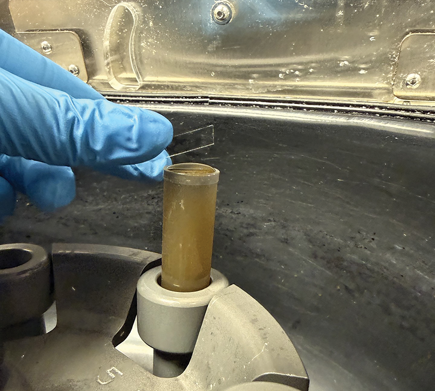
Figure 1. A veterinary team member places a coverslip on a centrifugal fecal flotation preparation with an appropriately sized reverse meniscus.
Deep Learning Algorithm
The available deep learning algorithm system (Vetscan Imagyst, Zoetis) has 3 components: a sample preparation device, a commercially available scanner, and analysis software.6-8 The results have been comparable to evaluations performed by diagnostic parasitologists using centrifugal or passive flotation techniques.8 This system has demonstrated acceptable identification of common gastrointestinal parasites in dogs and cats, including Giardia, Cystoisospora, Ancylostoma, Toxocara, and Trichuris species and Taeniidae eggs.6-8 The device is easy to use, requires minimal training for sampling preparation, and does not rely on a trained parasitologist for parasite identification.7
To date, deep learning algorithms depend on the information that has been added into the software and can accurately and consistently recognize a limited number of parasites. A visual examination of the slides supplements the use of deep learning algorithms when parasites are not correctly recognized or difficult to recognize (e.g., small protozoa).
Fecal Polymerase Chain Reaction
PCR testing is a highly sensitive technique that provides a convenient complement to traditional microscopic examination methods since it requires less feces and achieves a species-level identification.11 Some commercial fecal PCR tests can detect parasite DNA.11
The presence of a parasite’s DNA in stool does not necessarily imply an active infection since the genetic material can be derived from nonviable eggs, dead adults, or parasite fragments. PCR does not differentiate among parasite life stages and is not a test that can be routinely performed in-clinic since it requires special equipment and training. Additionally, a variety of factors may reduce the sensitivity of the test, such as intrinsic PCR inhibitors within feces or nonspecific primer binding.
Coproantigen Immunoassays
Coproantigen detection uses capture and detection antibodies to detect specific antigens from intestinal parasites, including roundworms (Toxocara, Toxascaris, and Baylisascaris species), hookworms (Ancylostoma and Uncinaria species), whipworms (Trichuris vulpis, Trichuris felis), flea tapeworms (Dipylidium caninum), Giardia species, and Cystoisospora species (Cystoisospora canis, Cystoisospora ohioensis-like, Cystoisospora felis, Cystoisospora rivolta).10,12-16 One commercial coproantigen test for Giardia species is able to differentiate life stages, and when it is used in conjunction with zinc sulfate centrifugal fecal flotation, more cases of Giardia infection are diagnosed.1,10 Coproantigen tests are validated for only a limited number of parasites, and most of the tests need to be sent to a referral laboratory.
Fecal Sedimentation
Eggs from trematodes, acanthocephalans, and some cestodes are heavier and do not float using flotation techniques; thus, sedimentation is the appropriate test.3 A total of 10 g of feces is mixed with 100 mL of water, which sits for an hour before the supernatant is decanted. The process is repeated until the supernatant becomes clear. A few drops of the sample from the bottom of the tube should be placed on a microscope slide, coverslipped, and read on the microscope.3
Baermann Test
The Baermann test can isolate larvae, including metastrongyloid lungworms, but the fecal sample used must be fresh and never frozen; otherwise, environmental nonparasitic soil nematodes can contaminate the sample.3 Larval identification requires advanced training in the minute differences in parasite morphology; therefore, confirmation through consultation with a veterinary parasitologist or PCR testing is recommended. Performing multiple Baermann tests increases the detection of larvae given the erratic pattern of larval shedding in lungworm infections.3
The Baermann test is performed by placing 10 g of feces in a double layer of cheesecloth, securing it beneath 2 applicator sticks using a rubber band, and suspending it in a water-filled funnel or a wine glass for 8 hours.3 A pipette is used to transfer the sediment at the bottom of the funnel to a glass slide for microscopic examination.3
Quantitative Fecal Examination Techniques
The advantages and limitations of currently available quantitative tests are briefly described below.
Modified McMaster
In small animal medicine, the modified McMaster technique is typically used in cases of suspected anthelmintic resistance to calculate eggs per gram (EPG) for use in the FECRT.3,17 Typically, a mixture of 4 g of feces with 56 mL (or 2 g of feces in 28 mL) of flotation solution (e.g., sodium nitrate) is used; alternatively, 4 g of feces can be mixed with 26 mL of flotation solution. After mixing, the mixture is strained and placed into both chambers of the McMaster slide, which sits for 5 minutes before reading (FIGURE 2).3 The slide should be read with the grid lines in focus, and eggs are counted at 10× magnification with each type of egg counted separately.
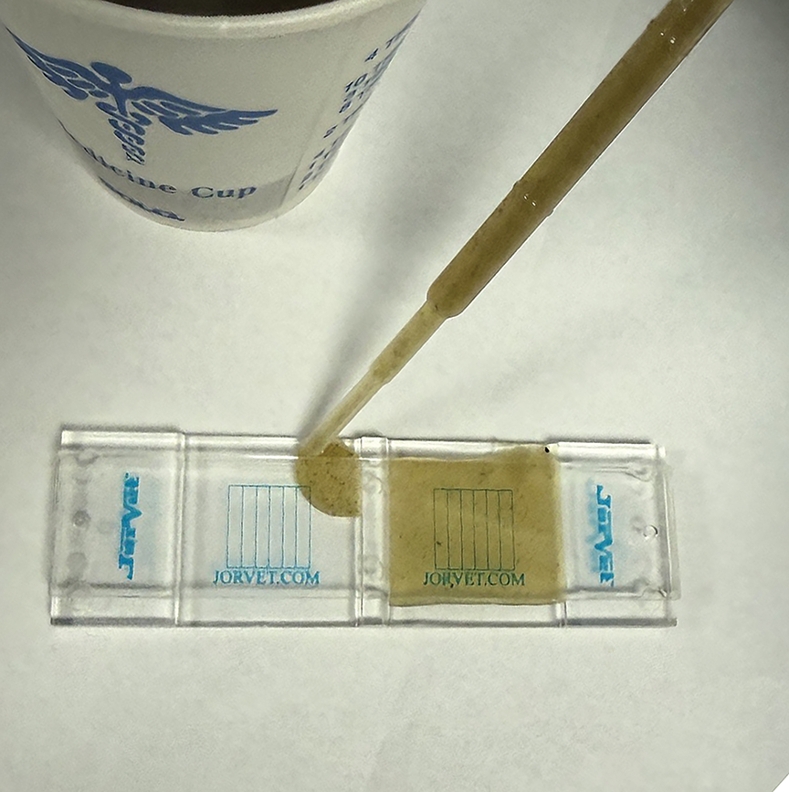
Figure 2. For the modified McMaster technique, a plastic disposable pipette is used to fill both sides of a McMaster slide with a prepared fecal solution containing animal feces and sodium nitrate solution.
To calculate the EPG, the egg count is multiplied by the appropriate conversion factor: 50 for mixtures containing 1 g of feces per 14 mL of solution or 25 for a mixture containing 1 g of feces per 6.5 mL of solution.3 It should be noted that this technique has a lower detection limit of 25 to 50 EPG (depending on the conversion factor), and the accuracy of this test is reduced when egg levels are below this limit of detection.3
Wisconsin
The Wisconsin egg count technique allows the quantification of no less than 1 EPG.3 The procedure is identical to the centrifugal flotation test, with the difference of recording the total weight of feces tested, which can be 1 to 5 g, and counting all parasite stages within the sample.3 The EPG for each parasite is calculated by dividing the total count by the number of grams used. Accuracy can be increased by mixing 5 g of feces with 22 mL of flotation solution (e.g., Sheather’s) and dividing the mixture into 2 tubes.3
Summary
Diagnosis of intestinal parasitic infections is essential to assess animal health, zoonotic risk, and disease transmission. Flotation methods have been the primary method of diagnosis for endoparasites; however, additional diagnostic methods, including coproantigen immunoassays, PCR, and deep learning algorithms, have been introduced into veterinary practice. It is fundamental that practitioners be familiar with the tests available and their advantages and limitations.
References
- Stafford K, Kollasch TM, Duncan KT, et al. Detection of gastrointestinal parasitism at recreational canine sites in the USA: the DOGPARCS study. Parasit Vectors. 2020;13(1):275. doi:10.1186/s13071-020-04147-6
- Sweet S, Szlosek D, McCrann D, Coyne M, Kincaid D, Hegarty E. Retrospective analysis of feline intestinal parasites: trends in testing positivity by age, USA geographical region and reason for veterinary visit. Parasit Vectors. 2020;13(1):473. doi:10.1186/s13071-020-04319-4
- Zajac AM, Conboy GA, Little SE, Reichard MV. Fecal examination for the diagnosis of parasitism. In: Veterinary Clinical Parasitology. 9th ed. Wiley-Blackwell; 2021:1-190.
- Bowman DD. Diagnostic parasitology. In: Georgis’ Parasitology for Veterinarians. 11th ed. Elsevier; 2020:349-454.
- Dryden MW, Payne PA, Ridley R, Smith V. Comparison of common fecal flotation techniques for the recovery of parasite eggs and oocysts. Vet Ther. 2005;6(1):15-28.
- Nagamori Y, Scimeca R, Hall-Sedlak R, et al. Multicenter evaluation of the Vetscan Imagyst system using Ocus 40 and EasyScan One scanners to detect gastrointestinal parasites in feces of dogs and cats. J Vet Diagn Invest. 2024;36(1):32-40. doi:10.1177/10406387231216185
- Kanski S, Busch K, Hailmann R, Weber K. Performance of the Vetscan Imagyst in point-of-care detection of Giardia duodenalis in canine fecal samples. J Vet Diagn Invest. 2025;37(1):63-70. doi:10.1177/10406387241279177
- Nagamori Y, Hall Sedlak R, DeRosa A, et al. Evaluation of the VETSCAN IMAGYST: an in-clinic canine and feline fecal parasite detection system integrated with a deep learning algorithm. Parasit Vectors. 2020;13(1):346. doi:10.1186/s13071-020-04215-x
- David ED, Lindquist WD. Determination of the specific gravity of certain helminth eggs using sucrose density gradient centrifugation. J Parasitol. 1982;68(5):916-919.
- Dryden MW, Payne PA, Smith V. Accurate diagnosis of Giardia spp and proper fecal examination procedures. Vet Ther. 2006;7(1):4-14.
- Hartmeyer GN, Hoegh SV, Skov MN, Dessau RB, Kemp M. Selecting PCR for the diagnosis of intestinal parasitosis: choice of targets, evaluation of in-house assays, and comparison with commercial kits. J Parasitol Res. 2017;2017:6205257. doi:10.1155/2017/6205257
- Burton KW, Michael H, Drake C. The utility of coproantigen testing in screening populations. Vet Parasitol. 2025;336:110459. doi:10.1016/j.vetpar.2025.110459
- Porter AL, Hanna R, Saucier J, et al. Analytical validation of an immunoassay for the detection of Cystoisospora spp. coproantigen in canine and feline fecal samples. J Vet Diagn Invest. 2026;38(1):130-134. doi:10.1177/10406387251385940
- Elsemore D, Bezold T, Geng J, Hanna R, Tyrrell P, Beall M. Immunoassay for detection of Dipylidium caninum coproantigen in dogs and cats. J Vet Diagn Invest. 2023;35(6):671-678. doi:10.1177/10406387231189193
- Elsemore DA, Geng J, Cote J, Hanna R, Lucio-Forster A, Bowman DD. Enzyme-linked immunosorbent assays for coproantigen detection of Ancylostoma caninum and Toxocara canis in dogs and Toxocara cati in cats. J Vet Diagn Invest. 2017;29(5):645-653. doi:10.1177/1040638717706098
- Elsemore DA, Geng J, Flynn L, Cruthers L, Lucio-Forster A, Bowman DD. Enzyme-linked immunosorbent assay for coproantigen detection of Trichuris vulpis in dogs. J Vet Diagn Invest. 2014;26(3):404-411. doi:10.1177/1040638714528500
- Jimenez Castro PD, Howell SB, Schaefer JJ, Avramenko RW, Gilleard JS, Kaplan RM. Multiple drug resistance in the canine hookworm Ancylostoma caninum: an emerging threat? Parasit Vectors. 2019;12(1):576. doi:10.1186/s13071-019-3828-6