
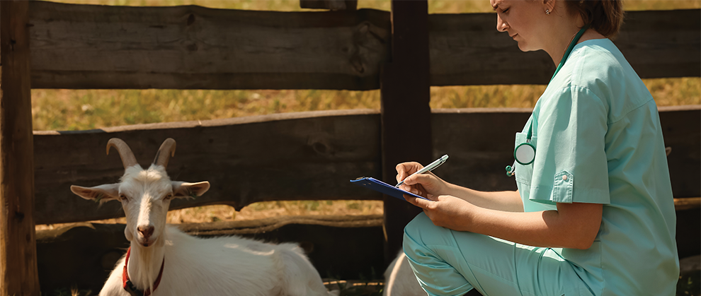
Neurologic diseases in goats account for approximately 5% of all cases submitted to diagnostic laboratories in the United States for necropsy. Despite this low percentage, clinicians are seeing more cases in practice, and adequate neurolocalization and diagnosis can be daunting. Neurolocalization requires an in-depth history, distance examination, and thorough physical examination before discussing diagnostics with the client (e.g., blood work, cerebrospinal fluid analysis, advanced imaging). Despite the wide range of differential diagnoses, treatment often includes supportive care with intravenous fluid therapy, rumen fluid transfaunation, thiamine supplementation, and antimicrobial administration.
Take-Home Points
- Adequate neurolocalization and diagnosis for goats often include taking a thorough history (e.g., clinical signs, medications administered by the client, vaccine status) and performing distance and physical examinations to build an adequate differential diagnosis list.
- Despite the wide range of differential diagnoses for neurologic disease, treatment often involves supportive care with oral or intravenous fluid therapy, rumen fluid transfaunation, thiamine supplementation, anti-inflammatory and antimicrobial therapy, and seizure control treatment as needed.
- Successful outcomes depend on neurolocalization, disease diagnosis, and time between onset of clinical signs and initiation of medical management.
Many conditions in goats have relatively pathognomonic clinical presentations that make the diagnostic process straightforward, but this is unfortunately not true of neurologic conditions. Although neurologic conditions account for only about 5% of all cases submitted to diagnostic laboratories for necropsy,1 the wide variety of clinical signs and the vast list of differential diagnoses make the clinical approach to the neurologic goat daunting. A comprehensive understanding of neurolocalization will help the clinician narrow down the list of neurologic differentials, and prioritizing the differential diagnoses based on frequency of presentation and pathognomonic findings will help the clinician select targeted diagnostic tests and treatment protocols to confidently manage goats with neurologic disease.
In working up a goat for neurologic disease, 2 findings will help ensure that the clinician does not overinterpret incidental findings unique to goats:
- Postmortem examination commonly reveals thalamic melanosis in brown-haired goats and occasionally in white-haired goats; this finding has no clinical significance.2
- The number and morphologic appearance of lumbar, sacral, and coccygeal vertebrae in goats vary (FIGURE 1).3,4 Up to 24% of goats possess 7 lumbar vertebrae, and the standard numbers of sacral and coccygeal vertebrae are usually reported as a range from 4 to 5 sacral and 10 to 12 coccygeal. The variable vertebrae are often referred to as transitional vertebrae because their morphologic appearance is between that of both sacral and coccygeal vertebrae.5 Awareness of these variations will help clinicians avoid overinterpreting radiographic images.
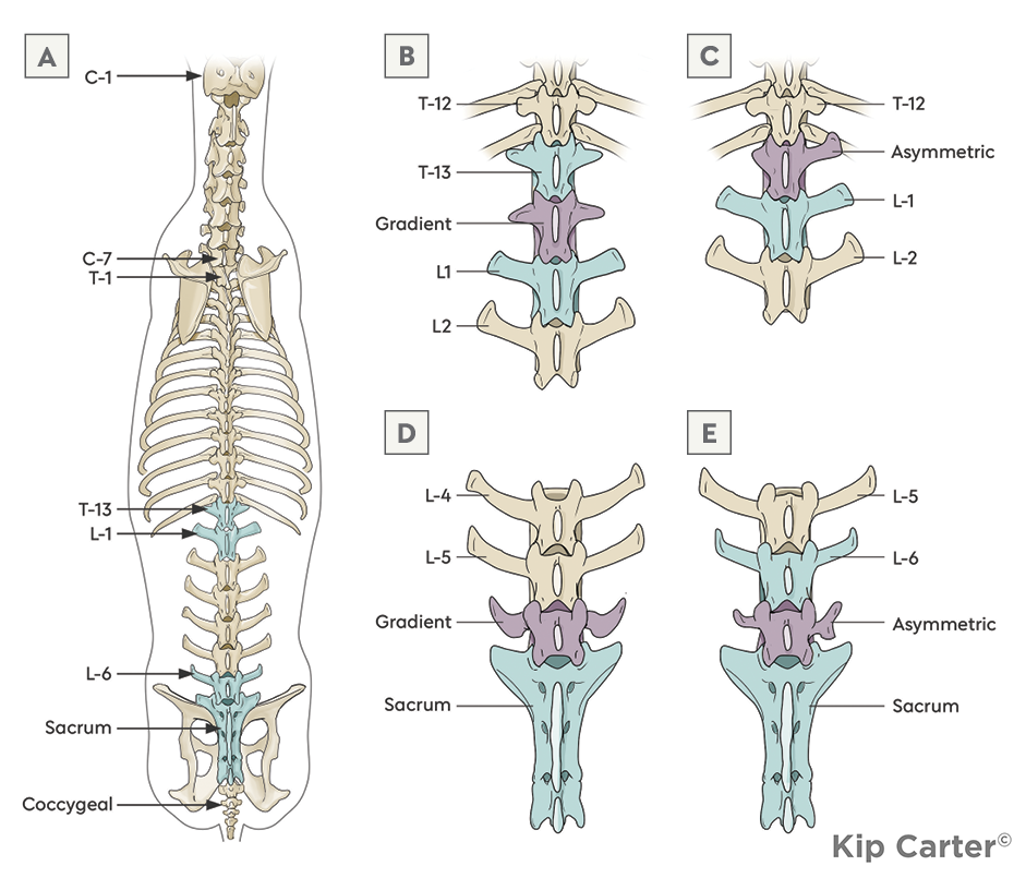
Figure 1. Anatomic variations of the axial skeleton of the goat. Transitional vertebrae (purple) can be referred to as gradient or asymmetric (blue highlights nonvariable neighboring vertebrae). Gradient vertebrae possess characteristics of the segments cranial and caudal to them (i.e., thoracic and lumbar) and are located at the junction between 2 segments of the skeleton. Asymmetric vertebrae can be better classified into 1 segment of the spinal column but demonstrate distinct left-to-right asymmetry in their lateral processes. (A) Traditional goat skeleton. (B) The most common location for a supernumerary gradient vertebra located at the thoracolumbar junction, demonstrating characteristics of both a thoracic and a lumbar vertebra. (C) The traditionally shaped T13 may be replaced by an asymmetric transitional vertebra that is missing a lateral process but articulates with a traditional first lumbar vertebra. (D) The transitional vertebrae may also be located at the lumbosacral junction, with the 6th lumbar vertebra displaying a morphology similar to the sacral shape but in the location of the anticipated L6. (E) L6 may be present as normal at the lumbosacral junction but, instead of articulating directly with the sacrum as seen in (A), it may articulate with a transitional vertebra, commonly displaying left-to-right asymmetry in the shape and size of the lateral processes. Illustration: Kip Carter
The nervous system comprises the brain, spinal cord, and peripheral nerves. Most commonly, when an animal is presented for neurologic disease, clients recognize pathology of the brain, brainstem, or spinal cord that result in changes in mentation, seizure activity, deficits in cranial nerves, incoordination, spinal ataxia, or inability to rise. Some of these presenting signs (e.g., seizures, cranial nerve deficits) are easy to localize to the neurologic system; however, others (e.g., ataxia, inability to rise) must be differentiated from musculoskeletal disorders. Performing a confident and informed neurologic examination is one of the mainstays for differentiating primary neurologic diseases from diseases of other body systems that result in similar clinical signs.
Because of the wide variety of purposes for which goats in the United States are raised and the growing popularity of goats as family pets, many clients are willing to pursue complete workups for advanced diagnostics, especially for patients exhibiting neurologic signs, as differential diagnoses may include zoonotic or herd management issues or both.
The Neurologic Examination
Any neurologic evaluation begins before the clinician places a hand on the animal. Learning the patient’s history and presenting sign are crucial to building a comprehensive differential diagnosis list; key features include duration and acuity of onset, herd size, number affected, and vaccination status. After a detailed history has been obtained, the clinician should observe the affected animal(s) from a distance without stimulation, which enables observation of mentation; interaction; willingness to eat; and, if eating, ability to chew and swallow food, eructate, and chew cud. After the distance examination, gently encouraging the affected animal to walk without a halter or assistance enables the clinician to evaluate what the animal is capable of and what it is readily willing to do.
Next, the authors prefer to perform a complete physical examination of the animal, ending with evaluation of the face, mouth, and cranial nerves, as handling even a well-socialized ruminant’s face may cause distress and elevate other clinical examination parameters. Even if there is an obvious neurologic deficit, a full physical examination including temperature, pulse, respiration, rumen auscultation, and evaluation of mucus membrane/FAMACHA (Faffa Malan Chart, a visual assessment scoring of conjunctival mucus membrane color in which a gradient of pallor indicates increasing severity of anemia) should be performed.6 A CBC and serum biochemistry may also provide key insights into systemic illness. A CBC enables clinicians to differentiate infectious diseases from traumatic processes and to rule out anemia leading to central hypoxia as a cause for neurologic impairment. Serum biochemistry enables clinicians to ensure that renal and hepatic function are normal before starting treatment. In addition, biochemistry parameters help clinicians rule out uncommon causes of neurologic diseases caused by decreased toxic waste clearance as well as metabolic/electrolyte abnormalities driving seizures, weakness, and recumbency; the presence of elevated muscle enzyme levels may support a diagnosis of trauma.
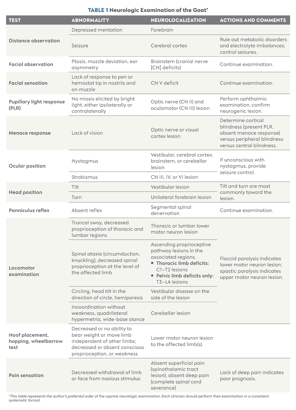
The neurologic examination is then broken down into segments: evaluation of mentation, cranial nerves, truncal reflexes and sensation, tail and anal tone and sensation, limb placement, conscious proprioception, and balance (TABLE 1). Unlike large ruminants and equine patients, goats and other small ruminants are able to undergo more complete evaluation of limb placement, hopping, and postural reaction evaluation similar to that conducted with small animals.
Differential Diagnoses
Most differential diagnoses can be subdivided into categories according to their primary neurolocalization (e.g., cerebral, cerebellar, brainstem, peripheral vestibular, spinal cord, peripheral neurologic, neuromuscular). A thorough neurologic examination will lead the clinician to determine which portion of the nervous system is affected, and other physical examination and blood work findings will help the clinician prioritize differential diagnoses within each neurolocalization (BOX 1).
- Central manifestation of peripheral/systemic disease
- Polioencephalomalacia
- Urea toxicity
- Hepatic encephalopathy
- Toxins
- Lead toxicity
- Salt toxicity/water deprivation
- Caprine arthritis encephalitis virus
- Trauma
- Skull fractures
- Thermal trauma from disbudding
- Neoplasia
- Rabies
- Aberrant parasitism
- Neosporosis
- Toxoplasmosis
- Parelaphostrongylus tenuis infection, in rare cases
- Bacterial meningitis
- Listeriosis
Differentials for neonates
- Hypoglycemia
- Hypoxia
- Bacterial meningitis
- Hydrocephalus/anencephaly
Cerebellum
- Cerebellar hypoplasia (usually secondary to congenital viral infection)
- Bluetongue
- Border disease
- Cache Valley virus infection
- Rift Valley fever
- Cerebellar abiotrophy
- Hereditary hypomyelinogenesis
- Grass staggers from Bermuda, rye, and Dallisgrass
- Neoplasia
Brainstem
- Listeriosis
- Bacterial meningitis
- P tenuis infection
- Neoplasia
Peripheral vestibular
- Otitis media
- Otitis interna
- Ear ticks
Spinal cord
- Trauma
- Neoplasia
- Enzootic ataxia (copper deficiency)
- Vertebral body abscess
- P tenuis migration
- Caprine arthritis encephalitis
- Toxic plant ingestion
- Lupinus (lupine or bluebonnet)
- Nicotiana species (flowering tobacco)
- Grass staggers
- Polyradiculoneuritis
Metabolic disease with neurologic manifestation
- Hypomagnesemia (grass tetany)
- Hypocalcemia (milk fever)
- Hypernatremia
- Hyponatremia
- Hypokalemia
*Neurolocalizations are ordered in the author’s opinion of frequency in which clinicians will encounter them.
After the clinician has determined a prioritized differential diagnosis list, further targeted diagnostic testing can be performed to confirm or rule out major differentials. Trauma is the one differential that can affect any location, and the clinical signs can vary significantly depending on the portion of the nervous system affected (VIDEO 1).
Polioencephalomalacia
Although polioencephalomalacia, also known as cerebrocortical necrosis, is one of the most commonly diagnosed central neurologic conditions of ruminants, it is a manifestation of other diseases, not a primary diagnosis. Polioencephalomalacia is caused by decreased thiamine (vitamin B1) levels, resulting from either absolute deficiency caused by inappetence and decreased feed intake or from increased thiaminase production caused by overgrowth of ruminal thiaminase-producing bacteria that leads to ruminal breakdown of thiamine and decreased systemic absorption. Thiamine is a key cofactor in the enzymatic reactions of glucose metabolism in the brain. Without enough thiamine, the brain becomes energy-deprived and the cerebral cortex undergoes necrosis.1,10
Clinical signs of polioencephalomalacia begin with tremors, ataxia, a staggering gait, and lethargy and progress to cortical blindness, head pressing, stargazing posture, recumbency, seizures, and death. The diagnostic workup for polioencephalomalacia often focuses on identifying or ruling out underlying causes of illness and thiamine depletion and includes a thorough history and individual physical examinations, including rumen sampling for microbial health and pH that would indicate clinically significant changes in feed intake.
Alternatively, polioencephalomalacia can be caused by increased sulfur intake, lead toxicity, or salt toxicity. Salt toxicosis can be ruled out via serum-to-cerebrospinal fluid (CSF) sodium ratios and lead toxicosis via serum or liver lead testing. In pregnant does, nervous ketosis should be ruled out with blood or urine ketones, and in young fast-growing kids, enterotoxemia may accompany convulsions, opisthotonus, and sudden death, which may be confused with polioencephalomalacia. Although supportive care and prompt provision of supplemental thiamine are essential for recovery, goats with polioencephalomalacia should be evaluated thoroughly and treated for the primary cause of the condition; otherwise, reoccurrence is common when thiamine supplementation is discontinued.
Listeriosis
Listeriosis is often one of the top differentials for neurologic disease in goats. It may share common clinical signs with polioencephalomalacia; however, clinical signs associated with brainstem abscesses, often presenting as cranial nerve deficits (e.g., facial nerve paralysis, ptosis, ear droop, head tilt/turn) as well as proprioception deficits (e.g., hypersalivation, inability to swallow or stand), are key hallmarks of listeriosis and help clinicians differentiate the 2 conditions (VIDEO 2).
Listeriosis is caused by the gram-positive bacteria Listeria monocytogenes, which can be found in soil, moldy hay, and silage and thrives in an environment with a low pH. Thus, it can affect a single animal or an entire herd. L monocytogenes is a bacterial infection that most commonly enters the central nervous system (CNS) from small abrasions in the oral cavity and is related to feeding with poorly fermented silage. The bacteria cause multifocal abscesses within the CNS as well as an associated diffuse meningitis. The bacteria have a predilection for the brainstem and thus often produce classic cranial nerve deficits; however, clinical signs may be less pathognomonic if abscesses appear in other cortices of the brain.11 The disease can be spread in aborted fetuses and placenta, feces, urine, and milk and is considered zoonotic to humans.
Prompt treatment with supportive care and antimicrobials is key, and clients should be made aware that reoccurrence is possible. Traditionally, listeria meningitis has been treated with either a penicillin or oxytetracycline, ideally administered intravenously to reach high serum concentrations rapidly. Although neither of these antibiotics has excellent CNS penetration in healthy animals, the presumption of blood–brain barrier breakdown in the presence of acute inflammation supports the clinical finding that these antibiotics are effective. Other antibiotics have been used off label with varying success, particularly trimethoprim-sulfonamides in preruminant-age goats as well as florfenicol. The rationale behind using other antibiotics is the increasing rate of antimicrobial resistance and higher rate of penetration of the blood–brain barrier.12,13
Caprine Arthritis Encephalitis
Although screening for the enzootic viral infection caprine arthritis encephalitis is common, the neurologic form is not as prevalent as polyarthritis and is therefore not as common a differential diagnosis as polioencephalomalacia and listeriosis. Although rare, most neurologic cases occur in juvenile animals with progressive spinal and central lesions.
Diagnostic Workup
For many patients, a thorough clinical examination and blood work are sufficient to lead to a diagnosis and treatment. If the diagnosis is not readily apparent or the clients are inclined to pursue further workup, advanced diagnostics may be dictated by the neurolocalization determined by the neurologic examination.
For most patients, the next diagnostic steps are more thorough investigation of trauma with diagnostic imaging or evaluating CSF via tap. Most goats are small enough that diagnostic head, neck, and spinal radiographs can be obtained by using a portable machine on the farm, although referral to facilities with advanced imaging and techniques (e.g., myelography) has become more common in recent years. CSF can be safely obtained from the lumbosacral space with the goat lightly sedated and restrained in sternal recumbency. For most goats, a 1.5-in, 20-g needle and a 3-mL syringe can be used after the lumbosacral area has been aseptically sterilized. Placing a small lidocaine bleb under the skin beside the intended vertebral space allows for increased patient comfort and decreased sedation. In large goats, a 3.5-in, 20-g spinal needle may be needed. The authors recommend taking a sample of no more than 1 mL CSF per 5 kg (11 lb) body weight. Most laboratories require a minimum of 1 mL CSF for complete analysis.
To perform a CSF tap, palpate a divot between 2 vertebral processes just caudal to a line drawn between the left and right ileal wings and insert the needle on midline with the bevel facing toward the head. Insert the needle until a pop is felt and gently aspirate to determine if the lumbosacral space has been entered. Normal CSF should be clear and colorless with a total protein less than 40 mg/dL and a cell count less than 3 cells/µL.1
For herd outbreaks of disease or situations in which a definitive diagnosis is needed (e.g., suspicion of a zoonotic disease), the most warranted course of action for acutely neurologic goats can be euthanasia and necropsy.
Treatment Approaches
Detailed descriptions of treatment protocols for each differential are outside the scope of this article; however, general approaches to neurosupportive care are outlined (TABLE 2). The mainstays of neurosupportive care are provision of anti-inflammatory therapy in the form of NSAIDs or steroids; provision of thiamine at 10 to 20 mg/kg IV or SC until the goat’s clinical status has improved; and antimicrobial therapy when indicated by presence of fever, elevated leukocyte count, or clinical signs consistent with listeriosis. Care should be taken when administering steroids in combination with NSAIDs, as doing so increases the risk for abomasal ulcer formation. Use of steroids is also contraindicated in pregnant animals due to the risk for abortion in early gestation. When selecting antimicrobials for the treatment of listeriosis, rising resistance to frequently used medications should also be considered.16
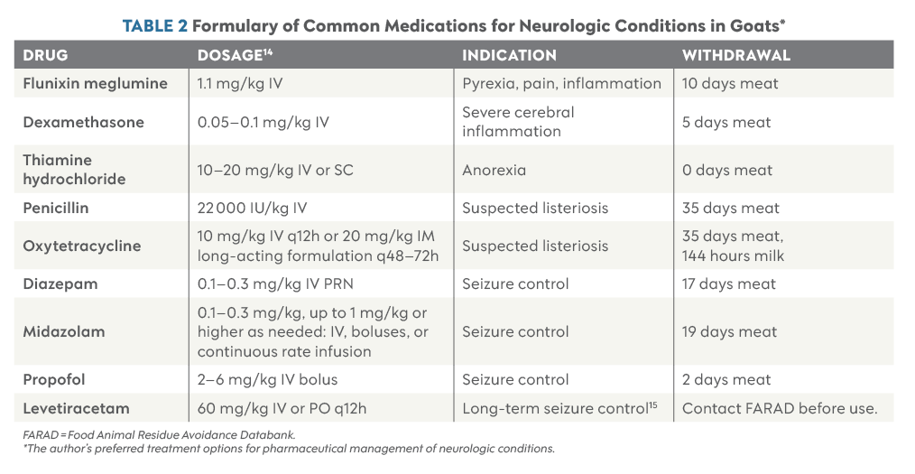
If animals are actively experiencing seizures, seizure control must be of the utmost priority, and animals that are unable or unwilling to eat and drink need hydration support in the form of intravenous balanced electrolyte solutions and thiamine support to prevent development of secondary polioencephalomalacia. In addition, if possible, anorectic goats with decreased rumen contractions should receive fresh rumen fluid transfaunation to support a normal ruminal microbial environment and further prevent development of polioencephalomalacia.
Summary
Although neurologic conditions among goats are relatively uncommon and the list of differential diagnoses is extensive, clinicians who take a systematic approach to examination and achieve accurate neurolocalization can successfully guide their clients toward well-informed diagnostic and treatment decisions.
Disclosure
Dr. Jamieson has received honoraria from Epicur Pharma for preparing and presenting continuing education content.
References
- Smith MC, Sherman DM. Nervous system. In: Smith MC, Sherman DM, eds. Goat Medicine. 2nd ed. Wiley-Blackwell; 2009:163-256. https://doi.org/10.1002/9780813818825.ch5
- Kaliner G, Frese K, Fatzer R, Fankhauser R. Thalamic melanosis in goats. Schweizer Archiv Fur Tierheilkunde. 1974;116(9):405-411.
- Simoens P, de Vos NR, Lauwers H, Nicaise M. Numerical vertebral variations and transitional vertebrae in the goat. Anat Histol Embryol. 1983;12(2):97-103. doi:10.1111/j.1439-0264.1983.tb01006.x
- Roels J, Hassoun R, Massenzio M, et al. A morphometric analysis of thoracolumbar vertebrae in goat by computed tomography. Vet Anim Sci. 2022;15:100233. doi:10.1016/j.vas.2022.100233
- Means KL, Zukeran-Kerr K, Le K, Yap SW, Brown K, Clarke L. Segmental megaesophagus secondary to extraluminal esophageal stenosis caused by transitional seventh cervical vertebra and supernumerary ribs in a goat. Vet Radiol Ultrasound. 2025;66(1):e70009. doi:10.1111/vru.70009
- Louge I, Lejuene M, Edwards M, et al. FaffaMalan Chart score is a poor single indicator for gastrointestinal parasitic burden of small ruminants in New York State. JAVMA. 2025;264(3):1-7. doi:10.2460/javma.25.07.0510
- Allen AL, Goupil BA, Valentine BA. A retrospective study of brain lesions in goats submitted to three veterinary diagnostic laboratories. J Vet Diagn Invest. 2013;25(4):482-489. doi:10.1177/1040638713493627
- Jones-Cook M. A practical approach to neurologic disease in small ruminants. Presented at: American Association of Bovine Practitioners Conference Proceedings; September 21-23, 2023; Milwaukee, Wisconsin. Accessed February 12, 2026. https://bovine-ojs-tamu.tdl.org/AABP/article/view/8895
- Nankemann J, Holland M, Glamann S, Cole R, Stockler J, Moon R. Spinal lymphoma in a goat. Vet Radiol Ultrasound. 2024;65(3):199-202. doi:10.1111/vru.13339
- Newsholme SJ, O’Neill TP. An outbreak of cerebrocortical necrosis (polioencephalomalacia) in goats. J S Afr Vet Assoc. 1985;56(1):37-38.
- Hu Y, Liu L, Zheng W, et al. Listeria monocytogenes invasion in goat brain tissues: mechanisms of blood–brain barrier disruption and regulation of apoptosis and autophagy. Front Microbiol. 2026;17:1748896. doi:10.3389/fmicb.2026.1748896
- Kennedy S, Passler T, Stockler J, Bayne J. Risk factors associated with outcome in goats with encephalitic listeriosis: a retrospective study of 36 cases from 2008 to 2021. J Vet Intern Med. 2023;37(3):1271-1277. doi:10.1111/jvim.16704
- López-Almela, I, Sheth CC, Gomis J, Gómez-Martin A, Lecit M, Quereda JJ. Epidemiology, clinical and pathological features and outcomes of listeriosis in ruminants: a systematic review and meta-analysis. Vet Q. 2025;45(1):2598257. doi:10.1080/01652176.2025.2598257
- The Ohio State College of Veterinary Medicine. Oxytetracycline [farm animal]. OSU VMC Antimicrobial Use Guidelines. Accessed February 12, 2026. https://ohiostate.pressbooks.pub/osuvmcabxuse/chapter/abx_oxytetracycline-farm-animal
- Stapley ED, Breuer RM Burton, AJ. Successful use of intravenous and oral levetiracetam in a goat to control refractory seizures secondary to suspected polioencephalomalacia. JAVMA. 2023;262(2):1-3. doi:10.2460/javma.23.09.0514
- Srinivasan V, Ham HM, Nguyen LT, Tamilselvam B, Murinda SE, Oliver SP. Prevalence of antimicrobial resistance genes in Listeria monocytogenes isolated from dairy farms. Foodborne Pathog Dis. 2005;2(3):201-211. doi:10.1089/fpd.2005.2.201