
Veterinarians in clinical practice often see patients that have vomiting as a clinical sign. As vomiting can have a wide range of etiologies, the cause can be challenging to diagnose. While many gastrointestinal parasites are primarily associated with diarrhea, vomiting may also be a key clinical sign of these infections. Parasitism is often overlooked as a cause of vomiting despite being relatively easy to diagnose and treat. This article provides key information and diagnostic strategies for practicing veterinarians about parasitic differentials for emesis in canine or feline patients.
Take-Home Points
- Vomiting is among the most common clinical presentations to the veterinary clinic and can have numerous causes, including gastrointestinal parasitism.
- Some parasitic infections can result in vomiting without any other clinical signs.
- Fecal diagnostic tests should be considered for all patients with vomiting of unknown etiology before more invasive or expensive diagnostic methods are used.
- Treatment and diagnostic strategies vary between different parasites, and no single diagnostic test can detect every parasite.
Vomiting is among the most common clinical signs observed in dogs and cats presenting to veterinarians and may result from a wide variety of underlying etiologies, such as various gastrointestinal disorders; dietary indiscretion; and infective agents, including parasites. Many of these diagnoses may be more common in dogs less than 1 year of age, when parasitism is also more commonly diagnosed. As a result, it can be challenging to work up cases of vomiting in pets. Diagnostic tests for common parasitic infections are cost-effective, minimally invasive tools that can help narrow the differential diagnosis.
The Value of Testing for Parasites
Many gastrointestinal parasites can cause vomiting, some even in prepatency. Parasites that cause vomiting include nematodes (roundworms), trematodes (flatworms), cestodes (tapeworms), and protozoa. In some cases, vomiting is an uncommon clinical sign, while in others it is the hallmark clinical sign and pets may present with no other abnormal findings.
Diagnostic tests for parasitic infections often require only a passively collected fecal sample or serial fecal samples for parasites with intermittent shedding. These tests can be performed in-clinic in less than 15 minutes, though some require specialized equipment, and reference laboratory options are commonly pursued. It is important to remember that detectability varies among parasite species, and a specific diagnostic test may not be the best option for every parasitic differential. For example, while centrifugal fecal flotation using a concentrated sugar solution is very effective at floating nematode eggs such as those of hookworms, roundworms, or whipworms, it may not be effective at floating heavier eggs such as Physaloptera or flatworm eggs, which are more effectively detected using fecal sedimentation.
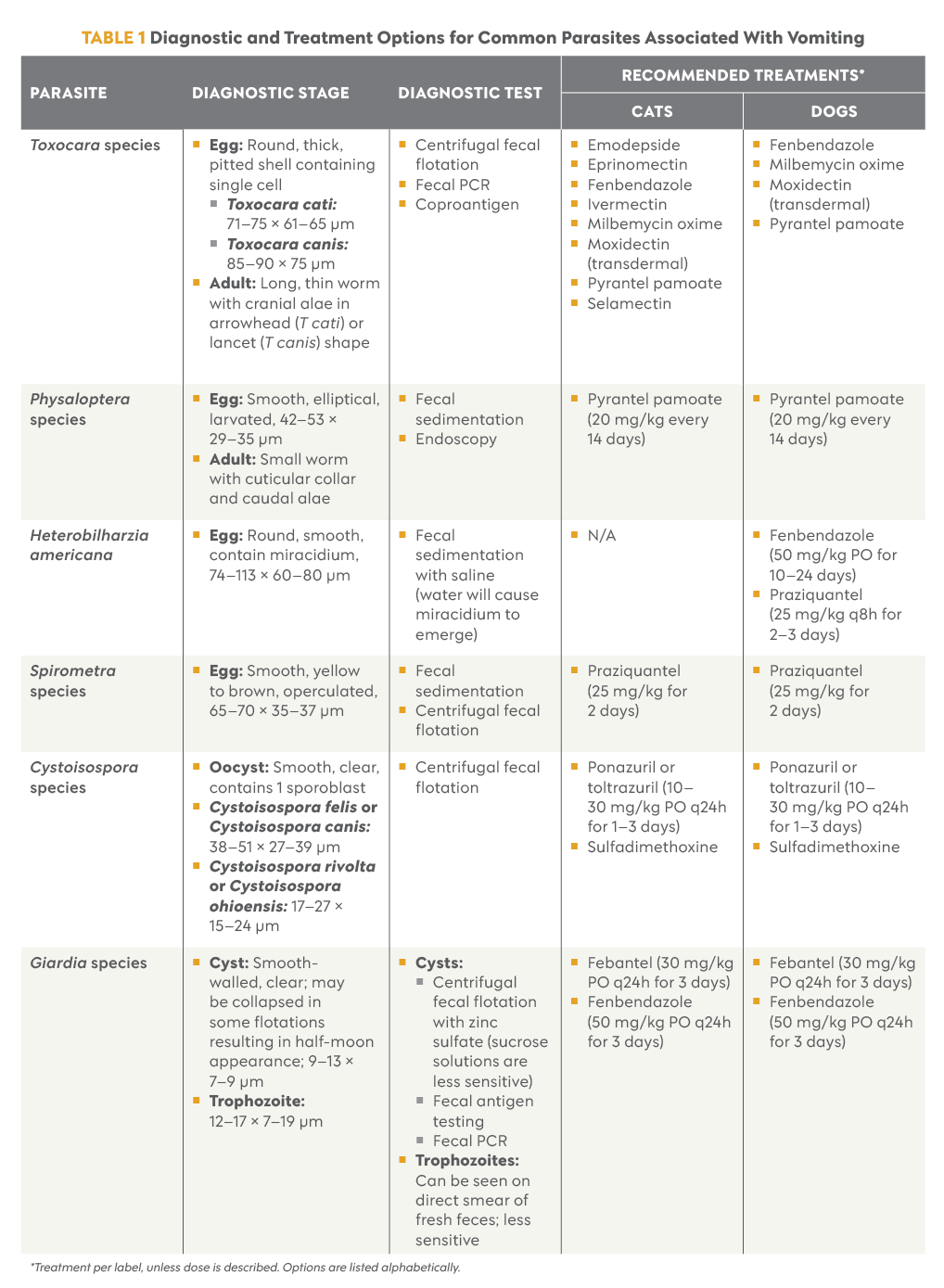
In cases of vomiting with no obvious life-threatening etiology (i.e., foreign body, toxin ingestion), quick and minimally invasive tests to rule out parasites should be performed before highly invasive or expensive diagnostics, such as exploratory laparotomy or endoscopic procedures, are considered. Table 1 summarizes diagnostic and treatment options for the most common parasites that should be considered in the differential list for a vomiting cat or dog.
Nematodes
Physaloptera species
Physaloptera species (Physaloptera rara and Physaloptera praeputialis) are spirurid nematodes that reside in the stomach of vertebrate hosts. Dogs and cats may serve as definitive hosts and are infected by ingesting arthropod intermediate hosts, typically crickets or beetles. Paratenic hosts may also be ingested by the definitive host.1,2
Clinical Signs
Vomiting is the hallmark clinical sign of Physaloptera infection, sometimes in the absence of any other clinical signs. Vomiting can occur even with low worm burdens and may persist long-term.3,4 These patients often do not have lethargy or inappetence but may be unable to keep food down.
Diagnosis
Eggs of Physaloptera are small and larvated (Figure 1). Typically, they are not detectable on regular centrifugal fecal flotation and require fecal sedimentation to be seen.5 Adults may be passed in vomitus and can be identified based on the presence of a cuticular collar and caudal alae.6
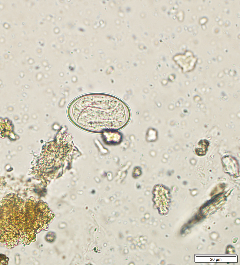
Figure 1. Physaloptera egg recovered via fecal sedimentation.
Toxocara canis and Toxocara cati
Toxocara species are roundworms that reside in the small intestine of the definitive host. T canis and T cati are very common parasites of puppies and kittens, respectively. Animals become infected with Toxocara species through multiple routes: after ingesting infective eggs from the environment, by consuming a paratenic host, or through transplacental transmission.7 Roundworms are extremely common, and nearly every litter will encounter them early in life.
Clinical Signs
Common clinical signs may include coughing (prepatent), vomiting (especially cats), and abdominal discomfort with a potbelly appearance. Severe disease is more often seen in young animals. If left untreated, severe infections can result in large masses of worms. Vomiting is more likely to be seen in cats than dogs.
Diagnosis
If roundworm infection is suspected, the ideal choice for in-clinic diagnostic testing is centrifugal fecal flotation using sugar solution.8 However, fecal PCR or coproantigen testing options exist for samples submitted to reference laboratories.9 Puppies and kittens should be tested frequently in the first year of life.
Trematodes
Heterobilharzia americana
H americana is a trematode parasite and the causative agent of canine schistosomiasis. When an infected dog or other mammal excretes feces contaminated with the parasite’s eggs, a larva (miracidium) emerges and infects freshwater snails (intermediate hosts). After some development in the snail’s tissues, the infective larval stage (cercaria) is released into water. Dogs become infected through skin penetration by these larvae when swimming in contaminated water.10
Clinical Signs
Most dogs are presented for veterinary evaluation due to chronic enteropathy. Clinical signs vary among patients and may include diarrhea, vomiting, weight loss, polyuria, polydipsia, fever, and dermatitis.10-12
Diagnosis
Diagnosis is achieved through a combination of tests, including fecal sedimentation, CBC, serum biochemical profile, urinalysis, and diagnostic imaging. The common clinicopathologic findings in dogs infected with H americana are anemia, eosinophilia, hypercalcemia or hypocalcemia, hyperglobulinemia, hypoalbuminemia, and isosthenuria. Fecal sedimentation for this parasite must use saline solution due to immediate hatching of the larvae when the eggs contact water (Figure 2).12,13
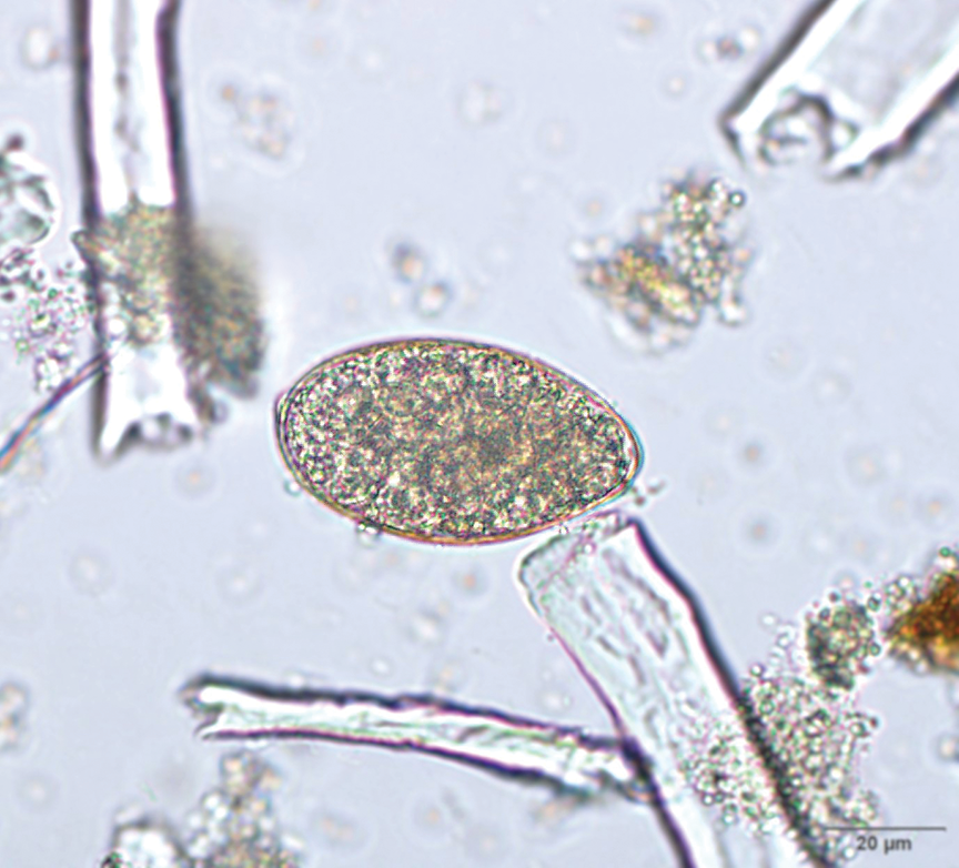
Figure 2. Heterobilharzia americana egg recovered from fecal sedimentation with saline.
Cestodes
Spirometra species
The adults of this cestode parasite infect the small intestine of dogs and cats, the definitive hosts. Eggs from infected animals are shed in water and subsequently release larvae called coracidia, which infect freshwater copepods. A larval stage called a procercoid develops in the infected copepods and later infects a second intermediate host—frogs, snakes, or any other small vertebrate except fish—where the final infective stage (plerocercoid) develops. Dogs and cats become infected when ingesting small vertebrates infected with the plerocercoid stage.14
Clinical Signs
Intestinal infections are usually asymptomatic, but in some cases, dogs and especially cats develop diarrhea, vomiting, lethargy, anorexia, and abdominal pain or distention. In some cats, proglottids can be seen in vomitus.15-17
Diagnosis
Fecal centrifugation with zinc sulfate or fecal sedimentation are the recommended diagnostic tests (Figure 3). Copro-PCR can help identify Spirometra species.14,17
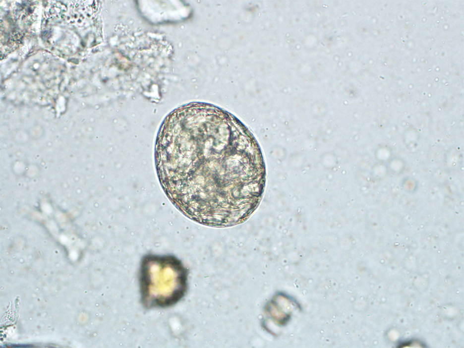
Figure 3. Spirometra egg recovered from fecal sedimentation.
Protozoa
Cystoisospora species
Infection occurs by ingestion of sporulated oocysts, either from the environment or in infected paratenic hosts or insects.18,19
Clinical Signs
Coccidiosis is commonly seen in puppies and kittens. Although the most common observation in infected animals is diarrhea, some animals present with a history of vomiting, lethargy, and abdominal pain.18,20,21
Diagnosis
Definitive diagnosis is made by observation of oocysts (Figure 4) during fecal centrifugal flotation in association with clinical signs.
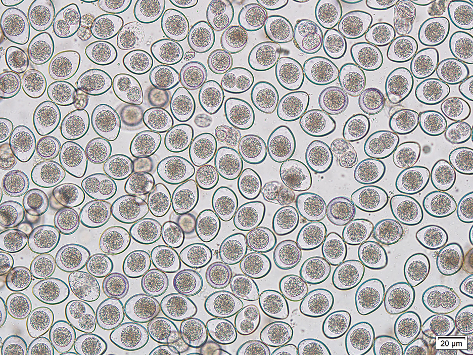
Figure 4. Numerous Cystoisospora oocysts recovered from fecal flotation.
Giardia species
Animals become infected by ingesting cysts, which are immediately infective once shed in the host’s feces.
Clinical Signs
Dogs and cats commonly have diarrhea, dehydration, and weight loss. Vomiting is occasionally observed.22
Diagnosis
Detection of Giardia cysts is achieved by centrifugal fecal flotation with zinc sulfate solution. Detection of trophozoites via direct smear is less common, as they do not survive in the environment as well as the cyst form does.
Other Parasites
Dirofilaria immitis, the causative agent of heartworm disease in dogs and of feline heartworm-associated respiratory disease, is a nematode parasite transmitted by mosquitoes, with the adult worms residing in the pulmonary arteries. Periodic vomiting is observed more commonly in cats than in dogs.23
Ollulanus tricuspis is a nematode parasite that lives in the stomach of cats and, rarely, dogs.24 Transmission occurs by ingestion of the infective larval stage present in vomitus of cats. The common findings in infected animals are chronic vomiting and gastritis. Diagnosis is made by Baermann testing of vomitus or gastric lavage fluid to recover parasitic larvae.25
Spirocerca lupi is a parasite found in esophageal nodules of dogs. It is transmitted by ingestion of intermediate hosts, primarily dung beetles, or of paratenic hosts such as rodents, rabbits, or chickens.26 In the United States, it has been reported mostly in wild canids from southern states.27 Infected dogs present with vomiting, chronic weight loss, dysphagia, and complications that include esophageal neoplasia.28 Diagnosis can be done by fecal sedimentation or centrifugal flotation with zinc sulfate to observe the larvated eggs, in combination with clinical signs and diagnostic imaging to detect nodules.
Oslerus osleri is a nematode parasite of dogs. Adults are located under the tracheal mucosa. In the United States, it has been mainly reported from coyotes. Transmission from dam to puppies occurs through saliva or feces containing the infective first larval stage. Clinical signs include coughing and vomiting. Diagnosis can be difficult but can be achieved by Baermann testing of feces or transtracheal wash samples. Bronchoscopy can also be utilized to detect tracheal nodules.29
Mesocestoides species are cestodes that infect the small intestine of dogs and cats. Infection is usually asymptomatic. On rare occasions, the parasite’s larvae penetrate the intestinal wall, leading to peritoneal larval cestodiasis. This condition can be severe and may cause a variety of clinical signs, including vomiting.30,31
Platynosomum fastosum is a trematode that parasitizes the bile ducts, gallbladder, and pancreas of cats. Infection occurs by ingestion of lizards, geckos, and other paratenic hosts. Clinical signs vary according to the number of adult parasites and can include vomiting, diarrhea, and icterus. In the United States, infection with this parasite is more common in southeastern states and Hawaii.32,33
Spurious Parasites
Dogs are naturally coprophagic, which can lead them to ingest parasites present in the feces of other hosts. Eimeria species are commonly identified in fecal centrifugation tests. Although spurious parasites are generally nonpathogenic, coprophagia can occasionally result in gastrointestinal signs such as vomiting.
Summary
Vomiting is a common presenting complaint encountered by general practice veterinarians. A wide range of etiologies can be responsible for vomiting in patients, including parasitism, which may sometimes be overlooked (CASE EXAMPLE). Various parasitic infections may cause vomiting in dogs and cats across the United States. A thorough understanding of the prevalence of these parasites, how animals get infected, treatment recommendations, and diagnostic strategies may be helpful for the practicing veterinarian to detect and treat parasites in the vomiting patient.
After 10 days of symptomatic care, clinical signs persisted and the owners elected to have an exploratory laparotomy performed. The duodenum was inflamed, and multiple 1-cm–long worms were found attached to the interior stomach wall. Toby’s veterinarian submitted these parasites, along with a fecal sample, to a reference laboratory for further examination.
Examination of the worms revealed that they were a species of Physaloptera, a stomach worm of dogs that commonly causes vomiting without other clinical signs.
What parasitic differentials could have been considered for Toby?
Of the parasites discussed in this article, Physaloptera should have been high on the suspicion list given the age of the patient and clinical presentation. Other parasites that could have been considered include Heterobilharzia, Spirometra, and Giardia, but further history probing could have helped rule out differentials.
What diagnostic test(s) could have been useful prior to surgical intervention?
Fecal diagnostic tests to rule out parasitism should have been performed, as these are fast and inexpensive. When Physaloptera is suspected, a fecal sedimentation test is advisable in addition to centrifugal flotation. Although surgery or endoscopy can detect Physaloptera, appropriate fecal diagnostics should be conducted first to reduce cost, time, and risk to the patient.
Would fecal diagnostic testing during the clinical manifestations have ensured a definitive diagnosis of Physaloptera infection?
Not necessarily. Use of an incorrect fecal diagnostic test, such as a regular passive or centrifugal flotation, will likely miss Physaloptera eggs. The best option is to perform fecal sedimentation to recover these eggs.
Prepatent infection can also cause false-negative results for Physaloptera. Even if these worms are immature and not yet producing eggs, their presence in the stomach can still cause irritation and clinical signs.
References
- Anderson RC. Order Spirurida – suborder Spirurina. In: Anderson RC, ed. Nematode Parasites of Vertebrates: Their Development and Transmission. 2nd ed. CABI; 2000:383-590. https://doi.org/10.1079/9780851994215.0383
- Lincoln RC, Anderson RC. Development of Physaloptera maxillaris (Nematoda) in the common field cricket (Gryllus pennsylvanicus). Can J Zool. 1975;53(4):385-390. https://doi.org/10.1139/z75-051
- Theisen SK, LeGrange SN, Johnson SE, Sherding RG, Willard MD. Physaloptera infection in 18 dogs with intermittent vomiting. JAAHA. 1998;34(1):74-78. doi:10.5326/15473317-34-1-74
- Soderman L, Harkin KR. Gastric Physaloptera infection in 27 dogs (1997-2019). JAAHA. 2021;57(1):8-14. doi:10.5326/JAAHA-MS-7046
- Kazacos KR. Just ask the expert: how do you detect Physaloptera species eggs? dvm360. October 1, 2010. Accessed July 30, 2026. https://www.dvm360.com/view/just-ask-expert-how-do-you-detect-physaloptera-species-eggs
- Naem S, Asadi R. Ultrastructural characterization of male and female Physaloptera rara (Spirurida: Physalopteridae): feline stomach worms. Parasitol Res. 2013;112(5):1983-1990. doi:10.1007/s00436-013-3356-9
- Overgaauw PAM, van Knapen F. Veterinary and public health aspects of Toxocara spp. Vet Parasitol. 2012;193(4):398-403. doi:10.1016/j.vetpar.2012.12.035
- Adolph C, Barnett S, Beall M, et al. Diagnostic strategies to reveal covert infections with intestinal helminths in dogs. Vet Parasitol. 2017;247:108-112. doi:10.1016/j.vetpar.2017.10.002
- Ascarid. Companion Animal Parasite Council (CAPC). Updated February 28, 2025. Accessed May 12, 2026. https://capcvet.org/guidelines/ascarid
- Graham AM, Davenport A, Moshnikova VS, et al. Heterobilharzia americana infection in dogs: a retrospective study of 60 cases (2010-2019). J Vet Intern Med. 2021;35(3):1361-1367. doi:10.1111/jvim.16127
- Rodriguez JY, Camp JW, Lenz SD, Kazacos KR, Snowden KF. Identification of Heterobilharzia americana infection in a dog residing in Indiana with no history of travel. JAVMA. 2016;248(7):827-830. doi:10.2460/javma.248.7.827
- Baniya A, Goldy CJ, Ardpairin J, et al. Canine schistosomiasis in the West Coast: Heterobilharzia americana in two natural intermediate hosts found in the Colorado River, California. Pathogens. 2024;13(3):245. doi:10.3390/pathogens13030245
- Aicher K, Poellmann L. Diagnosis and treatment of Heterobilharzia americana infection in dogs. Todays Vet Pract. 2025;15(2):24-29.
- Rojas A, Salazar-Grosskelwing E, Beck R. Spirometra mansoni. Trends Parasitol. 2026;42(5):1471-4922. doi:10.1016/j.pt.2026.01.001
- Kim J, Ock Y, Yang K, et al. First clinical cases of spirometrosis in two cats in Korea. Korean J Parasitol. 2021;59(2):153-157. doi:10.3347/kjp.2021.59.2.153
- Laing S. Canine Spirometra infection. The University of Sydney Centre for Veterinary Education. December 2022. Accessed August 11, 2026. https://www.cve.edu.au/common/Uploaded%20files/CT/CANINE-SPIROMETRA-article.pdf
- Alvarado-Hidalgo I, Campos-Camacho J, Arguedas-Morales Y, et al. Molecular, morphological and histopathological evidence of Spirometra mansoni in wild and domestic animals from Costa Rica. Vet Parasitol Reg Stud Reports. 2024;51:101030. doi:10.1016/j.vprsr.2024.101030
- Buehl IE, Prosl H, Mundt HC, et al. Canine isosporosis – epidemiology of field and experimental infections. J Vet Med B Infect Dis Vet Public Health. 2006;53(10):482-487. doi:10.1111/j.1439-0450.2006.00973.x
- Mitchell SM, Zajac AM, Charles S, Duncan RB, Lindsay DS. Cystoisospora canis Nemeseri, 1959 (syn. Isospora canis), infections in dogs: clinical signs, pathogenesis, and reproducible clinical disease in beagle dogs fed oocysts. J Parasitol. 2007;93(2):345-352. doi:10.1645/GE-1024R.1
- Morelli S, Di Cesare A, Traversa D, et al. Occurrence of Cystoisospora spp. and other intestinal parasites in dogs and cats with diarrhea. Vet Parasitol. 2025;338:110546. doi:10.1016/j.vetpar.2025.110546
- Dubey JP, Lindsay DS. Coccidiosis in dogs—100 years of progress. Vet Parasitol. 2019;266:34-55. doi:10.1016/j.vetpar.2018.12.004
- Kurnosova OP, Panova OA, Arisov MV. Prevalence of Giardia duodenalis in dogs and cats: age-related predisposition, symptomatic, and asymptomatic cyst shedding. Vet World. 2024;17(2):379-383. doi:10.14202/vetworld.2024.379-383
- Litster AL, Atwell RB. Feline heartworm disease: a clinical review. J Feline Med Surg. 2008;10(2):137-144. doi:10.1016/j.jfms.2007.09.007
- Kato D, Oishi M, Ohno K, et al. The first report of the ante-mortem diagnosis of Ollulanus tricuspis infection in two dogs. J Vet Med Sci. 2015;77(11):1499-1502. doi:10.1292/jvms.15-0158
- Cecchi R, Wills SJ, Dean R, Pearson GR. Demonstration of Ollulanus tricuspis in the stomach of domestic cats by biopsy. J Comp Pathol. 2006;134(4):374-377. doi:10.1016/j.jcpa.2006.02.003
- Rojas A, Dvir E, Baneth G. Insights on Spirocerca lupi, the carcinogenic dog nematode. Trends Parasitol. 2020;36(1):52-63. doi:10.1016/j.pt.2019.10.004
- Alfaro-Segura P, Robleto-Quesada J, Montenegro-Hidalgo VM, et al. Elucidating Spirocerca lupi spread in the Americas by using phylogenetic and phylogeographic analyses. Front Parasitol. 2023;2:1249593. doi:10.3389/fpara.2023.1249593
- Mylonakis ME, Rallis T, Koutinas AF, et al. Clinical signs and clinicopathologic abnormalities in dogs with clinical spirocercosis: 39 cases (1996–2004). JAVMA. 2006;228(7):1063-1067. doi:10.2460/javma.228.7.1063
- Conboy G. Helminth parasites of the canine and feline respiratory tract. Vet Clin North Am Small Anim Pract. 2009;39(6):1109-1126. doi:10.1016/j.cvsm.2009.06.006
- Carta S, Corda A, Tamponi C, et al. Clinical forms of peritoneal larval cestodiasis by Mesocestoides spp. in dogs: diagnosis, treatment and long term follow-up. Parasitol Res. 2021;120(5):1727-1735. doi:10.1007/s00436-021-07107-w
- Venco L, Kramer L, Pagliaro L, Genchi C. Ultrasonographic features of peritoneal cestodiasis caused by Mesocestoides sp. in a dog and in a cat. Vet Radiol Ultrasound. 2005;46(5):417-422. doi:10.1111/j.1740-8261.2005.00076.x
- Haney DR, Christiansen JS, Toll J. Severe cholestatic liver disease secondary to liver fluke (Platynosomum concinnum) infection in three cats. JAAHA. 2006;42(3):234-237. doi:10.5326/0420234
- Xavier FG, Morato GS, Righi DA, Maiorka PC, Spinosa HS. Cystic liver disease related to high Platynosomum fastosum infection in a domestic cat. J Feline Med Surg. 2007;9(1):51-55. doi:10.1016/j.jfms.2006.08.006