
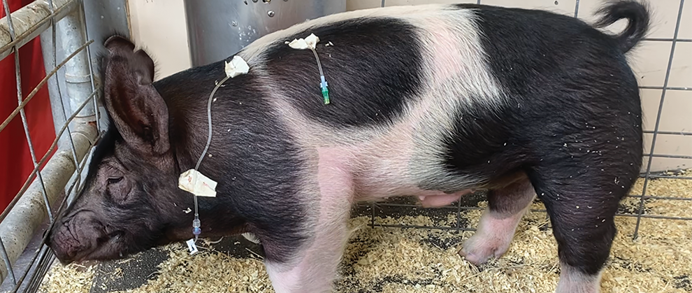
Although potbellied pigs are called “food animals” for regulatory purposes, they are increasingly being acquired as companion animals. A common medical concern for potbellied pigs is vomiting, which can be secondary to surgically treated conditions (e.g., obstruction) or medically treated conditions (e.g., gastrointestinal stasis, ulceration). This article will discuss the diagnostic approach to a pig that is vomiting, including physical examination, hematology, and imaging. In addition, common causes—including gastrointestinal obstruction, stasis, and ulceration—will be reviewed along with treatment options.
Take-Home Points
- The initial diagnostic plan for a vomiting potbellied pig is similar to one for a vomiting dog or cat and includes collecting a history, performing a physical examination, submitting a CBC and serum biochemical profile, and performing abdominal radiography.
- The most common causes of vomiting among potbellied pigs are gastrointestinal obstruction, stasis, and ulceration.
- Less common causes of vomiting among potbellied pigs include postoperative pain, infectious agents, toxins, and neoplasia.
- Gastrointestinal obstruction and stasis can be differentiated by radiography.
- Ulceration can be confirmed via fecal occult testing or gastroscopy.
- Fluid therapy for pigs can be challenging due to patient compliance. Some pigs may tolerate rectal fluid therapy, which enables fluid administration without the challenge of acquiring intravenous access.
Potbellied pigs are deemed “food animals” for regulatory purposes, yet they are increasingly being acquired as companion animals. Unfortunately, resources about pigs housed in urban environments as companion animals remain sparse. This article will discuss the clinical approach to a potbellied pig that is vomiting, a common medical concern of this species.
The most common causes of vomiting among pigs include gastrointestinal obstruction, stasis, and ulceration as well as postoperative pain, infectious agents (e.g., influenza, transmissible gastroenteritis, salmonellosis), toxins, and neoplasia.1,2 General gastroenteritis is not a common cause of vomiting among pigs, unlike in dogs. A thorough diagnostic approach should be taken to determine the cause of vomiting on initial examination rather than merely treating the clinical signs. After the cause of vomiting is identified, the treatment plan is often clear and direct. Early intervention typically results in a favorable prognosis.
Because pigs are legally classified as food animals, regardless of their status as companion animals, meat withdrawal times must be discussed and documented for all medications used for every porcine patient, even if the client does not plan to use the patient for food consumption. Federal guidelines must be followed for extralabel drug use, and the Food Animal Residue Avoidance Databank should be consulted to establish appropriate withdrawal intervals for medical records. Adherence to these regulations is vital for food safety and the protection of one’s veterinary professional license.
Initial Diagnostic Plan
The initial diagnostic plan for a pig that is vomiting is remarkably similar to a plan for a dog or cat that is vomiting. A thorough history should be collected, followed by a physical examination, CBC, serum biochemical profile, and radiography with or without a barium study.
Collecting a History
The collection of a thorough and accurate history may be crucial to an efficient, effective diagnostic plan. Depending on a pig’s environment, a client may directly state that the pig is vomiting. However, if the pig is housed outside or in a barn stall–type setting, a client may state that the pig has a poor appetite. A client should be asked about any changes to the diet and environment (e.g., housing, addition of pets) as well as the likelihood of getting into trash and access to toys. Additional questions should be asked about exposure to healthy and sick animals or humans. Questions about the frequency and timing (i.e., immediately after eating, hours later) of defecation and vomiting as well as consistency of feces and vomitus should also be asked. The answers to these questions will determine the list of differential diagnoses. For example, if a pig has recently consumed trash or destroyed a toy, gastrointestinal obstruction will be a more likely cause for vomiting. If a pig has experienced a sudden stressor or diet change, gastrointestinal stasis or ulceration may be a more likely cause. Finally, if a pig has been exposed to a sick animal or human, an infectious agent may be a more likely cause.
Blood Analysis
A CBC and serum biochemical profile should be performed to discern the next appropriate steps in the diagnostic and treatment plan. A pig with gastrointestinal obstruction will most likely have a stress leukogram paired with metabolic alkalosis, hypochloremia, hypokalemia, and dehydration.3 A pig with gastrointestinal ulceration may have regenerative anemia with elevated blood urea nitrogen and low total protein.4,5 A pig with an infectious cause for vomiting will have hematologic inflammatory changes and possibly low albumin and hypocalcemia with dehydration.5 These hematologic changes are similar to those displayed by other companion animal species, such as dogs and cats, that are vomiting.
Imaging
Imaging is necessary to determine if a pig is obstructed and to identify the location of the obstruction. Abdominal radiography is considered the standard of care for a pig that is acutely vomiting. A barium study may also be indicated to determine if an exploratory laparotomy is necessary. The radiographic changes that indicate gastrointestinal obstruction are similar to those noted among dogs and cats. If radiography is inconclusive, computed tomography may be necessary.
Sedation may be required to ensure human and pig safety while good-quality diagnostic radiographs are acquired. However, care needs to be taken to protect airway access, as anesthesia drugs are known to increase the risk for vomiting. Aspiration of barium and/or vomit into the lungs must be prevented. Diazepam is an option for sedation because it has not been shown to increase the risk for vomiting.6 The use of α2-agonists may increase the risk for vomiting and should be avoided.7
Fluid Therapy
Regardless of the cause of vomiting, fluid therapy is the mainstay of supportive care. The kind of fluid used for pigs is similar to that used for other species and depends on hydration status and acid–base levels as determined by hematology. Isotonic fluids are key to rehydration. Fluids may be administered orally, intravenously, rectally, intraperitoneally, or intraosseously, depending on the condition of the pig.
Oral fluids are the simplest route of administration; however, oral consumption can be difficult, especially for a pig in a hospitalized environment. Many oral electrolyte formulations are commercially available for pigs.
Intravenous fluid rates have been reported from 1 mL/kg/hr to 6 mL/kg/hr, and 2 mL/kg/hr is typically reported as a maintenance rate.3,8 Intravenous access can be difficult to obtain, but common locations are the lateral or central auricular vein, superficial epigastric vein, jugular vein (requires a cut down), cephalic vein, and medial or lateral saphenous vein.9 A useful resource for intravenous fluid therapy that includes catheter location followed by recommended catheter size has been published elsewhere.9
Fluids may be administered rectally if a pig is not in critical condition. Continuous rectal fluid administration may be done by attaching a red rubber catheter to the tail. One study compared rectal fluid therapy at a rate of 4.4 mL/kg/hr (i.e., more than double the maintenance fluid rate) to intravenous fluid therapy, and the results suggested that this rectal fluid rate may not be sufficient to replace intravenous fluids.8 One study has shown that horses become agitated and have increased fecal water content when administered fluids at higher rates (e.g., 5 mL/kg/hr).10 To the author’s knowledge, higher fluid rates have not been investigated in the pig. Thus, rectal fluid therapy may be provided when venous access is unavailable but should not be relied on to support rehydration.
Fluids may be administered intraperitoneally or intraosseously as a last resort for rehydration for a dehydrated pig in critical condition. With these routes of fluid therapy, the objective is to stabilize the patient then transition to intravenous fluids. Intraosseous catheterization can be performed at the humerus or trochanteric fossa of the femur yet may require special equipment depending on the age of the pig.9
Causes of Vomiting
Obstruction
The history may contain pertinent details that suggest gastrointestinal obstruction is the likely cause of vomiting (e.g., got into the trash, ate corn cobs, destroyed a toy). Typically, if the onset of vomiting was relatively recent, a pig’s mentation will be bright and alert. As time progresses and vomiting continues, mentation will deteriorate. Body temperature and lung sounds are typically within normal limits. Feces may be firm or absent depending on the chronicity of obstruction. If the obstruction is at the level of the stomach, gastrointestinal endoscopy or gastrotomy is warranted.
Gastrointestinal endoscopy can be performed if the obstructing item is within the stomach, has blunt edges, and is small enough to move retrograde through the esophagus. If the obstructing item is within the small intestine, an enterotomy, and possibly resection and anastomosis, is warranted. The surgical approach for a gastrotomy or enterotomy is along the ventral midline. One difference in the anatomy of pigs, in comparison to other species, is that the mesenteric arteries are in bundles. During a resection, care and precision are required to ensure each intestinal section has proper blood supply via mesenteric bundles. Postoperative complications include incisional dehiscence (internal and/or external), infection, and devascularization. Postoperative pain management, fluid therapy, gastroprotectants, and antibiotics are warranted if a gastrotomy or enterotomy is performed.
Gastrointestinal Stasis
The diagnostic approach to gastrointestinal stasis is similar to that used for obstruction. The 2 pathologies can be differentiated by radiography. For patients with gastrointestinal stasis, radiography will indicate a feces-filled large intestine with no evidence of an obstructive pattern/intestinal obstruction. Understanding the cause of gastrointestinal stasis is crucial for successful treatment. Potential causes include environmental stress, pain (including postoperative pain), dehydration, or postprocedural (e.g., tusk trimming, feet trimming) resulting from a combination of stress and medications (e.g., sedatives, anesthetic agents). The treatment will be determined by the cause. Appropriate treatment to correct the cause—such as adequate pain management, fluid therapy, or prokinetic drug—is required.
Gastrointestinal Ulceration
Gastrointestinal ulceration is often multifactorial and among the more commonly studied causes of vomiting in pigs. It is typically induced by physiologic stress or diet and can be confirmed by a fecal occult test. In addition, gastroscopy can be used to confirm the diagnosis and determine the size and location of ulceration, if necessary.
If gastric ulceration is confirmed, the diet of a pig should be adjusted to increase dietary fiber (i.e., nonpelleted or mashed feedstuff), which may be done by adding leafy greens, pumpkin, or potato starch to the diet.1 Proton pump inhibitors are also recommended at a dose of 40 mg PO q24h.11 Mucosal protectants (e.g., sucralfate) may be used as a component of supportive care but may not be used as a solitary treatment. Because of their short half-life, histamine receptor antagonists have been shown to be ineffective for pigs, especially if not administered via intravenous catheter.12 A treatment plan that addresses the underlying cause (e.g., stress, diet) is more likely to be effective at treating the patient and preventing future recurrence.1
Other Causes
Other causes of vomiting for potbellied pigs are infectious agents and toxins, yet these are less likely causes, especially for a pig in a single-pig household. These causes are typically ruled out by the history and imaging along with inflammatory hematology findings, fecal PCR, and feed toxin screening tests. The treatment for infectious and toxin-mediated causes of vomiting is primarily supportive care according to a pig’s clinical signs, which is most likely fluid therapy for dehydration and potentially maropitant to minimize vomiting.12,13
Although gastrointestinal obstruction is more likely a result of dietary indiscretion, neoplasia—resulting in obstruction of the intestinal lumen—has also been reported. Intestinal carcinoma is one of the more common types of neoplasia identified in potbellied pigs.14
Summary
The diagnostic and treatment plans for a potbellied pig that is vomiting is similar to plans for a dog or cat that is vomiting. A thorough history should be collected, and a physical examination should be performed. Hematology and diagnostic imaging will further determine the treatment plan. Common causes include gastrointestinal obstruction, stasis, and ulceration. The treatment plan typically includes fluid therapy, diet modification, and gastroprotectants. Surgery and antimicrobials may be prescribed as needed. Appropriate meat withdrawal times must be considered due to the pig’s classification as a food animal species (regardless of companion animal status) within the United States.
References
- Mozzachio K. Illness and disease. In: Mozzachio K, ed. Potbellied Pig Veterinary Medicine. 1st ed. Elsevier; 2023:129-212.
- Ludwig EK, Byron CR. Evaluation of the reasons for and outcomes of gastrointestinal tract surgery in pet pigs: 11 cases (2004–2015). JAVMA. 2017;251(6):714-721. doi:10.2460/javma.251.6.714
- Hobbs KJ, DeNotta SL, Gallastegui A, et al. Obstipation in pet pigs: 24 cases. Can Vet J. 2021;62(8):843-848.
- Anderson LC, Mitchell J, Ringler DH, Bohr DF. Cimetidine prophylaxis for gastric ulcers in laboratory swine. Lab Anim Sci. 1981;31(5):498-501.
- Clark SG, Coffer N. Normal hematology and hematologic disorders in potbellied pigs. Vet Clin North Am Exot Anim Pract. 2008;11(3):569-582. doi:10.1016/j.cvex.2008.03.003
- Szelenyi I, Herold H, Göthert M. Emesis induced in domestic pigs: a new experimental tool for detection of antiemetic drugs and for evaluation of emetogenic potential of new anticancer agents. J Pharmacol Toxicol Methods. 1994;32(2):109-116. doi:10.1016/1056-8719(94)90062-0
- Smith JS, Seddighi R. Miniature companion pig sedation and anesthesia. Vet Clin North Am Exot Anim Pr. 2022;25(1):297-319. doi:10.1016/j.cvex.2021.08.007
- Chigerwe M, Blasczynski SJ, Abi-Nader BA, Condy PM, Kretsch CM, Depenbrock SM. Continuous fluid infusion per rectum compared with intravenous fluid infusion in pigs. J Vet Intern Med. 2023;37(4):1580-1587. doi:10.1111/jvim.16733
- Elane GL, Bauck AG, Hobbs KJ, et al. Review of venipuncture and intravenous catheterization techniques in pigs. JAVMA. 2024;262(10):1388-1396. doi:10.2460/javma.24.03.0169
- Khan A, Hallowell GD, Underwood C, van Eps AW. Continuous fluid infusion per rectum compared with intravenous and nasogastric fluid administration in horses. Equine Vet J. 2019;51(6):767-773. doi:10.1111/evj.13113
- Friendship RM, Melnichouk SI, Dewey CE. The use of omeprazole to alleviate stomach ulcers in swine during periods of feed withdrawal. Can Vet J. 2000;41(12):925-928.
- Doster AR. Porcine gastric ulcer. Vet Clin North Am Food Anim Pract. 2000;16(1):163-174. doi:10.1016/s0749-0720(15)30141-9
- Smith JS, Gebert JE, Ebner LS, et al. Pharmacokinetics of intramuscular maropitant in pigs (Sus scrofa domesticus). J Vet Pharmacol Ther. 2023;46(3):158-164. doi:10.1111/jvp.13120
- Newman SJ, Rohrbach B. Pot-bellied pig neoplasia: a retrospective case series (2004-2011). J Vet Diagn Invest. 2012;24(5):1008-1013. doi:10.1177/1040638712452725