
To take the CE quiz, click here.
This quiz is open until April 2028.
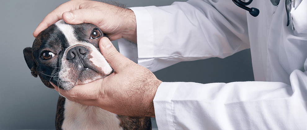
Brachycephalic patients, with their charming expressions and juvenile features, are at risk for a variety of ocular problems resulting from their appearance and anatomy. Excessively long eyelids, shallow orbits, eyelid conformation, and lash abnormalities—along with a variety of other anomalies (e.g., tear film deficiencies, corneal hypoesthesia)—predispose these patients to chronic ocular exposure, which can lead to discomfort and globe- or vision-threatening sequelae.
Take-Home Points
- Facial conformation has an undeniable impact on the health of the ocular surface, and brachycephalic patients have a greater risk for conditions that threaten the clarity and integrity of the cornea.
- Risk factor recognition and client education are critical to the management and mitigation of brachycephalic ocular syndrome.
- An excessively long eyelid and shallow orbit make a globe more susceptible to proptosis.
- Deviating or superfluous hairs (e.g., distichiasis, ectopic cilia, trichiasis) can be irritating and initiate or exacerbate corneal disease.
- Corneal ulcers may progress rapidly in brachycephalic patients and should be addressed promptly and assertively.
- Early recognition of ocular surface disease and early intervention to improve the anatomic protective functions of the adnexa can help maintain vision and comfort.
In addition to enduring the well-recognized brachycephalic obstructive airway syndrome (BOAS), brachycephalic dogs and cats also experience a related syndrome of ophthalmic abnormalities that can predispose them to discomfort and vision-compromising sequelae. Humans are drawn to the endearing, neotenous features of the short-faced, big-eyed brachycephalic dog, so much so that the French bulldog is the most popular breed of dog in the United States for the fourth year in a row according to the American Kennel Club.1 However, those big eyes with their winsome qualities can be problematic for these breeds. They typically have long palpebral fissures that do not provide adequate coverage of their prominent globes in addition to shallow orbits and other features that decrease globe protection. Add a spectrum of suboptimal factors such as aberrant eyelid and hair orientation, tear film deficiencies, and reduced corneal sensation to these features, and an unhealthy ocular surface flourishes.
Components of Brachycephalic Ocular Syndrome
Brachycephalic ocular syndrome may affect dogs and cats, yet the ubiquitous popularity of brachycephalic dog breeds compared to the niche popularity of brachycephalic cat breeds results in dogs being more commonly affected. The components of this syndrome exist on a spectrum and not all patients will be similarly or severely affected; however, the clinician should be aware of the following components.2
Euryblepharon
Euryblepharon, also known as macroblepharon, is an abnormally long eyelid opening or excessive length to the palpebral fissure (FIGURE 1). Most dogs do well if their palpebral fissures are approximately 6 to 8 mm longer than their corneal diameter; however, brachycephalic patients may have markedly longer fissures.3,4 An excessively long eyelid leads to inadequate apposition with the globe and the potential for globe exposure and dessication. In many cases, even if these dogs can complete an effective blink, their lids cannot close completely (lagophthalmos) or frequently enough to lubricate and protect the ocular surface. In addition, scleral show, or exposed white of the eye, is associated with a markedly increased risk for corneal ulcer development in brachycephalic patients.5 Even with a mere 10% increase in relative eyelid aperture width, the risk for corneal ulceration more than triples.5

Figure 1. Classic appearance of brachycephalic ocular syndrome in a pug. Note the enlarged palpebral fissure that allows excessive “show” of the sclera, poor coverage of the cornea, trichiasis from the medial canthus and a nasal fold, and tear staining present nasal and ventral to the eye. There is also evidence of chronic ocular surface irritation with corneal fibrosis and extensive vision-compromising superficial pigment.
Lagophthalmos
Lagophthalmos, an incomplete or inefficient blinking mechanism, can disrupt ocular surface homeostasis and increase the risk of developing exposure keratitis from reduced globe protection and impaired tear admixing and distribution. Most dogs with macroblepharon experience lagophthalmos to some degree, but even dogs without excessively long palpebral fissures can have an ineffective blink, especially if they experience facial paralysis or paresis or if they have any degree of neurologic or cognitive dysfunction. A recent study showed that lagophthalmos was present in 82% of Shih Tzus, with an estimated degree of incomplete eyelid closure ranging from 5% to 35% (average 15%).6 Other brachycephalic breeds may be similarly affected.
Medial Canthal Entropion
The lower eyelid, adjacent to the medial canthus, rolls inward and covers up, or occludes, the lower nasolacrimal puncta (FIGURE 2). This results in epiphora with the potential for moist facial dermatitis, particularly in animals with medial facial folds. This anatomic configuration frequently results in or exacerbates medial trichiasis.

Figure 2. The normal anatomic arrangement of the eyelid margins and cilia relative to the surface of the globe and various abnormal variations. Ectopic cilia: the lash is exiting through the palpebral conjunctiva and coming in direct contact with the cornea. Trichiasis: normal facial hairs are oriented toward and contacting the cornea. Entropion: the eyelid margins are inverted permitting dermal hair to contact the corneal surface. Distichia: the lash is exiting the tarsal gland ductal opening and is directed toward the ocular surface. Illustration: Kip Carter
Nasolacrimal Duct Malformation
A consequence of the foreshortened head conformation of brachycephalic breeds, the nasolacrimal duct can be grossly malformed. Frequently, their ducts are short and their canaliculi are elongated, and the course of the entire system can have an anomalous inverted U- or V-shaped appearance.7 In addition, these patients consistently have accessory openings into the posterior nasal cavity, resulting in fluid drainage away from the nasal vestibule.7 The shape and steep slope of their ducts and accessory openings do not necessarily cause clinical signs unless they exist concurrently with obstructive lesions. Nonetheless, they can change the results of the Jones test (fluorescein passage) assessing patency with either extended times to dye appearance or absence of dye at the nares if it drains into the nasopharynx and/or oropharynx.
Trichiasis
Hair that deviates toward the cornea from the medial caruncle, medial lower eyelid, or nasal skin folds adjacent to (and sometimes contacting) the cornea is called trichiasis (FIGURE 2).8 (The medial caruncle is a raised nodule of conjunctiva overlying glandular tissue that sits in the corner of the eye adjacent to the medial canthus.) This hair chronically irritates the ocular surface—even if the patient does not demonstrate discomfort—and can contribute to keratitis and ocular melanosis (superficial pigment migrating from the medial limbus toward the axial cornea).9
Distichiasis and Ectopic Cilia
Distichia are normal cilia that arise from follicles in the tarsus and emerge from the lid margin through meibomian gland openings (FIGUREs 2 AND 3). Distichia may or may not be irritating, depending on if they contact the cornea surface and their degree of stiffness.

Figure 3. Distichiasis of the upper and lower eyelid of a brachycephalic mixed-breed dog. Also note the trichiasis from the medial facial skin.
Ectopic cilia protrude directly through the palpebral conjunctiva. Ectopic cilia are typically along the upper eyelid at the 12 o’clock position; they often result in discomfort because the emerged hairs rub against the cornea with every movement of the upper eyelid (FIGURES 2 AND 4).
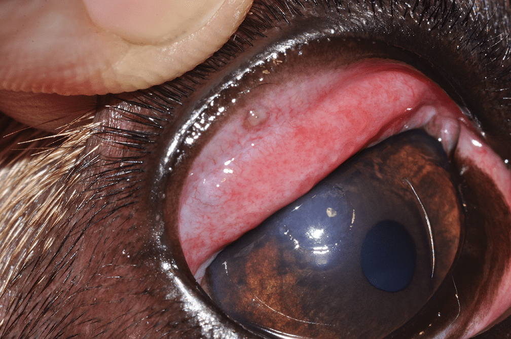
Figure 4. An ectopic cilia protruding from the dorsal bulbar conjunctiva corresponds to a dorsal paraxial corneal ulcer in a young French bulldog.
Inconsistent blepharospasm and recurrent or nonhealing corneal ulcers, sometimes in a vertical orientation, are the classic clinical signs of distichiasis and ectopic cilia. Brachycephalic breeds, especially Shih Tzus and English bulldogs, are the most frequently and severely affected.10
Reduced Corneal Sensitivity
The anterior corneal stroma and epithelium have a dense concentration of sensory nerve endings, which serve to alert the animal to damaging stimuli and initiate reflex tearing and normal healing processes.4 Brachycephalic patients may not exhibit acute or severe corneal pain, but diminished innervation can contribute to delayed recognition of a problem. Ongoing damage may lead to impaired corneal wound healing.
Quantitative or Qualitative Tear Deficiencies
The tear film is an admixture of lipid produced by the meibomian glands of the eyelids, an aqueous fraction produced by the lacrimal glands, mucin produced by conjunctival goblet cells, and glycocalyx mucin produced by corneal epithelial cells.
Quantitative dry eye is very common in brachycephalic patients and happens when the quantity of liquid tears is decreased relative to reference ranges. Aqueous tear secretion, as determined by a Schirmer tear test, is often lower in brachycephalic dogs compared with nonbrachycephalic dogs, including brachycephalic dogs without complaints of ophthalmic disease.11 This is likely multifactorial in origin and may be attributed in part to the reduced corneal sensitivity of brachycephalic patients and its negative effect on the afferent pathway of lacrimation.11,12 The stimulation of nasal mucosa via breathing accounts for approximately one-third of basal tearing in humans; thus, it has been proposed that abnormal airflow through the nasal cavity of dogs with BOAS contributes to reduced tear production in these canine breeds.11
Qualitative dry eye happens when the aqueous fraction of the tear fluid is of normal volume but the lipid and/or mucin fractions are deficient. This can be demonstrated by retention of the ocular stains rose bengal or lissamine green, which attach to degenerating cells and foci on an ocular surface that is inadequately covered by mucin (FIGURE 5). Brachycephalic breeds frequently have a significantly lower tear film breakup time—a diagnostic test that is an indirect measure of the mucin and lipid layers of tear film—than nonbrachycephalic breeds.2 Impaired meibomian gland function (also known as meibomian gland dysfunction) and increased ocular exposure predispose brachycephalic dogs to a significantly thinner lipid layer and diminished tear film stability, thereby increasing the risk for functional keratoconjunctivitis sicca.13 Thus, quantitative or qualitative tear deficiencies can cause or exacerbate corneal injuries and inflammation.
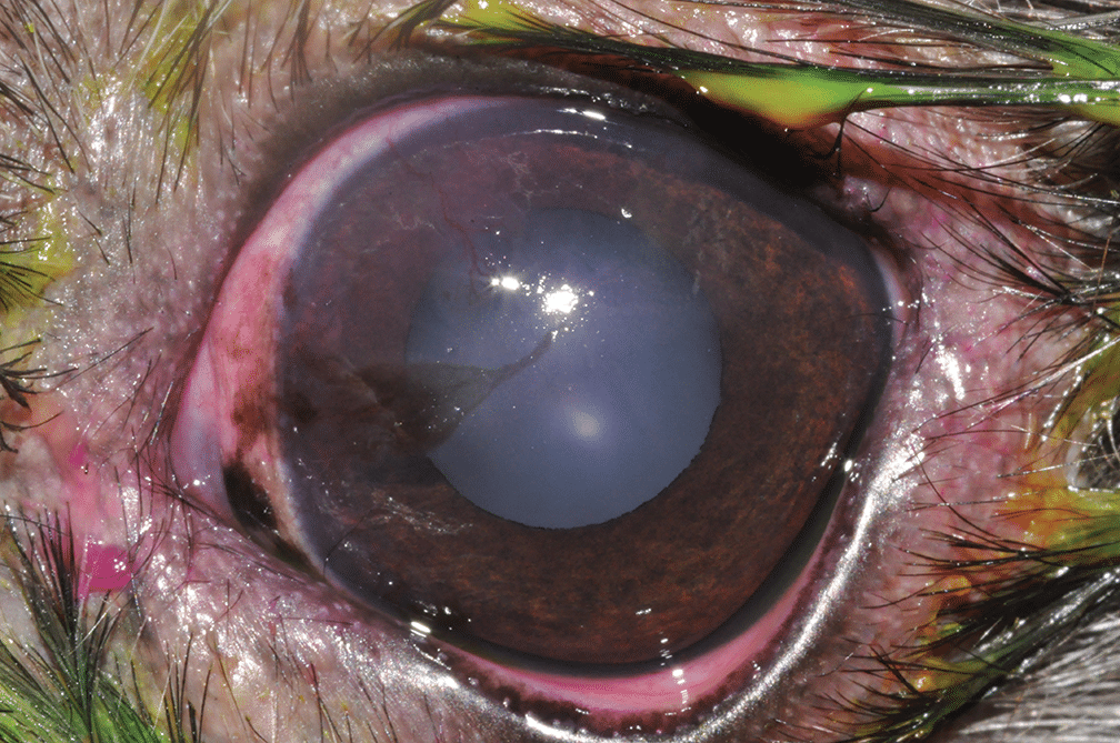
Figure 5. This Shih Tzu exhibits medial canthal entropion and trichiasis and has developed pigmentary keratitis. This is exacerbated by a qualitative tear film deficiency demonstrated by retention of rose bengal stain and superficial corneal vasculature.
Shallow Orbits
Brachycephalic breeds have a distinctive skull structure with shortened facial bones and thickened retrobulbar soft tissue, which contributes to a greater degree of ocular protrusion.14 Their shallow orbits provide considerably less globe protection than the deeper orbits of their mesocephalic or dolichocephalic counterparts. This orbital anatomy, especially when combined with excessively long palpebral fissures, greatly increases the potential for traumatic proptosis, a globe-threatening condition in which the eyelid margins become entrapped behind the equator of the globe, making it impossible for the patient to blink and protect the anterior structures of the globe.15 Damage to the optic nerve and extraocular muscles is common upon the initial injury. If the condition is not promptly repaired, corneal exposure and ulceration may ultimately lead to globe rupture.
Cherry Eye
Prolapse of the nictitans gland, also colloquially known as “cherry eye,” results from weakening of the connective tissue that normally keeps the nictitans gland tucked securely around the base of the interior cartilage, which gives the nictitating membrane its shape and structure. This permits migration of the gland above and forward of the membrane’s margin. Exposure of the gland results in inflammation and hypertrophy. Brachycephalic breeds have a considerably greater (nearly 7 times the odds) risk for prolapse of the nictitans gland compared with breeds that have mesocephalic skull conformations.16
Ocular Consequences
Brachycephalic ocular syndrome can result in myriad consequences.
Epiphora and tear staining result from inappropriate tear fluid drainage that, sometimes, is significant enough to cause moist dermatitis ventral to the eye.
Exposure keratitis is a broad term that encompasses the effects of impaired physical and functional coverage of the corneal surface. When the corneal surface desiccates, chronic inflammation results in the ingrowth of corneal vasculature, migration of superficial pigmentation, and damage to the corneal stromal extracellular matrix leading to fibrosis—all of which compromise the clarity of the visual axis.
Pigmentary keratitis is characterized by progressive pigmentation (i.e., melanosis) of the corneal surface, often combined with various degrees of corneal vascularization, scarring, and edema. This condition is common in brachycephalic breeds, and most common in pugs.17 A study of pugs found that there is a significant association between medial entropion of the lower eyelid and presence of pigmentary keratitis in this breed.17 In addition, severity of medial entropion of the lower eyelid is associated with severity of pigmentary keratitis, which worsens over time.17 Pigment changes sometimes form a whorl pattern initially but are most prominent in areas with the greatest degree of contact irritation from trichiasis, notably along the medial cornea (FIGURE 6).

Figure 6. Early medial corneal pigment in a pug accompanied by corneal vasculature. There is prominent scleral show, medial entropion, and medial trichiasis.
Corneal ulcers are lesions in which there is loss of corneal epithelial barrier and varying degrees of underlying stroma. Any single component of brachycephalic ocular syndrome can contribute to the development or exacerbation of a corneal ulcer, and it is well established that brachycephalic dogs have increased risk for ulcer formation compared to their mesocephalic and dolichocephalic counterparts.18 Unfortunately, brachycephalic dogs are also more likely to have deeper corneal involvement when they have an ulcer.19 Physiologic exophthalmos and impaired blinking most likely contribute to a thinner lipid layer and central decrease in the aqueous layer of tear film. Any compromise to the barrier function of the ocular surface increases the possibility of pathogen adherence and invasion, resulting in contamination and complicated ulcerative keratitis (FIGURES 7 AND 8). Often, surgical stabilization of deep or perforating corneal ulceration is necessary in these patients.

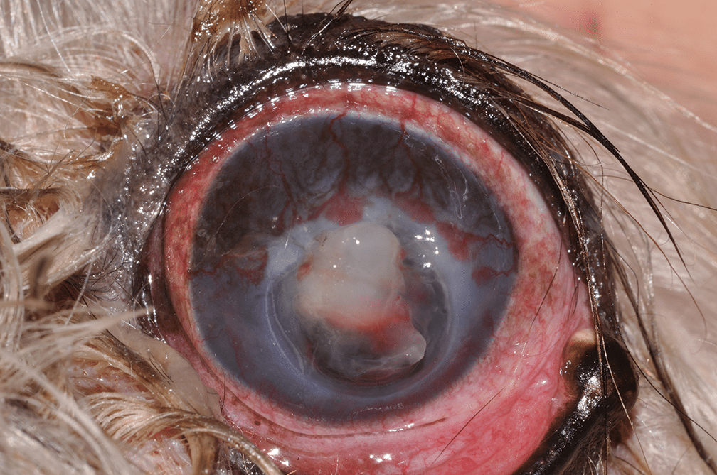
Proptosis results when the eyelid margins become entrapped behind the equator of the globe, preventing blinking.15 Euryblepharon, combined with a shallow orbit, favors this condition. A considerable amount of force is not needed to cause proptosis in a brachycephalic animal compared to animals with better globe protection. Although this condition is more frequent in brachycephalic breeds, the prospect for globe retention and, in some instances, retention of vision is greater. However, the longer the globe is exposed and unprotected, the greater the likelihood of corneal ulceration, which may progress to globe rupture. Proptosis is an emergency. In brachycephalic dogs, rapid repair can salvage vision as well as retain a comfortable globe (FIGURE 9).

Figure 9. Proptosis in a Shih Tzu. The eyelid margins are not visible because they have become entrapped posterior to the equator of the globe. The epithelium can be seen peeling away from the underlying stroma.
Squamous cell carcinoma is rare, but chronic corneal exposure and inflammation—especially if combined with topical immunosuppressive therapy or ultraviolet radiation—can result in epithelial dysplasia, which in rare instances transforms into squamous cell carcinoma. Brachycephalic breeds have a strong breed predilection for primary corneal squamous cell carcinoma (77%), particularly those prone to keratoconjunctivitis sicca (FIGURE 10).20

Figure 10. Corneal squamous cell carcinoma in a Shih Tzu with chronic dry eye receiving long-term topical cyclosporine.
Treatments and Preventive Strategies
Medical Options
Tear Supplements
Dry eye—qualitative and quantitative—results in an increase of tear electrolytes, proteins, and inflammatory mediators, which can damage the tissue of the ocular surface. One of the best symptomatic strategies to support the ocular surface is to supplement with artificial tears, which lubricate and dilute those factors that can be damaging at higher-than-normal concentrations. Unfortunately, the benefits of lubrication are short-lived and frequent administration is recommended. Tear supplements that contain hyaluronic acid (sodium hyaluronate), especially cross-linked hyaluronic acid, are superior for moisture and retention. Those containing carboxymethylcellulose or methylcellulose add viscosity, which has a longer duration of effect. The more viscous the product, the longer its surface contact time and action. For this reason, gels and ointment preparations are recommended when practical, especially before an anticipated extended period without treatment (e.g., when an owner leaves for work, prior to bed).2
Lacrimostimulants
Most cases of keratoconjunctivitis sicca in dogs have an immune-mediated etiopathogenesis in which lymphocytic infiltration with subsequent damage to the lacrimal glands impairs their function.11,12 Lacrimostimulant medications, specifically the calcineurin inhibitors such as cyclosporine and tacrolimus, prevent T-cell proliferation and differentiation. These drugs are useful not only for quantitative dry eye but have been shown to increase tear quality and decrease ocular surface inflammation. In addition, these drugs are commonly used for keratitis and do not have the same degree of negative adverse effects as topical corticosteroids.
Surgical Options
Eyelid Shortening Surgery
To determine ideal eyelid fissure size, start with an idea of what a dog’s ideal eyelid length would be, then plan to make both palpebral fissures that same length. The average dog’s corneal diameter is approximately 14 to 16 mm, but the palpebral fissure length may range from 23 to 38 mm (even longer in some dogs).3,4 Most dogs do well if their palpebral fissures are approximately 6 to 8 mm longer than their corneal diameter. However, this is merely a guideline. The perfect eyelid fissure size for a dog depends upon its anatomy and desired cosmetic outcome. Blepharoplasty procedures involve a degree of aesthetic awareness and understanding of the function of the eyelid.
Medial Canthoplasty
Medial canthoplasty is a technique that addresses many of the features of brachycephalic ocular syndrome in a single procedure.3,4,21-24 In addition to shortening the overall length of the fissure, this technique also removes medial caruncular tissue, which is often a source of irritating hairs, and corrects medial inferior entropion, which relieves the functional obstruction of the nasolacrimal apparatus. This procedure requires precision to identify and avoid the nasolacrimal punctal openings and canaliculi during tissue excision and suturing, remove the medial caruncular and conjunctival hair follicles, and appose the medial canthus.
Nasal Fold Resection
If hairs along the nasal facial folds contact the medial cornea, resection of this redundant tissue should be considered. Dogs with nasal folds are nearly 5 times more likely to be affected by corneal ulcers than those without.5 In this procedure, the fold is lifted and excised with large scissors and the resultant wound closed primarily.4 The appearance of the face will be altered; thus, clear communication with the client regarding expectations is recommended. This surgery will not address palpebral fissure length, caruncular trichiasis, or medial entropion.
Removal of Distichiasis/Ectopic Cilia
Deviated lashes can cause significant discomfort and predispose corneal disease. These offending hairs should be removed; singular or a few hairs can be surgically excised. Ectopic cilia should be surgically excised. Primary therapy for distichia and adjunctive therapy following ectopic cilia excision may include cryoepilation, especially for patients with a plethora of hairs.4,21
Monitoring and Other Management Strategies
Ophthalmic Examination
This should be a part of an annual examination—especially for breeds predisposed to keratoconjunctivitis sicca, older patients, and dogs with ocular signs (e.g., discharge, conjunctivitis, keratitis)—and include a Schirmer tear test-1. This examination can identify at-risk patients and enable early intervention. For every additional year of life, the risk for the development of keratoconjunctivitis sicca or corneal pigmentation increases by 10% and the risk for the development of meibomian gland dysfunction (qualitative tear deficiency) increases by 20%.12
Regular Grooming
Consistent trimming of periocular hair can be a helpful prophylactic step to minimize contact with the ocular surface.2 Cleaning and drying periocular hair and the spaces beneath the facial folds as part of a consistent hygiene routine gives caretakers the opportunity to observe the appearance of the eyes and notice, hopefully early, changes that may be concerning.
Early Referral
When facial conformation is extreme or ocular complications have already developed, consider referral to an ophthalmologist for anatomic correction (i.e., medial canthoplasty and nasal fold resection) and removal of irritating cilia. In addition, early consultation with a specialist for patients with complicated ocular surface disease, especially those with infected corneal ulcers, may help avoid the rapidly deteriorating corneal breakdown that these patients can experience. Surgical stabilization of deep corneal wounds can be vision- and globe-sparing.
Breeding and Welfare Consultations
Encourage breeders to avoid breeding animals with extreme brachycephalic conformations, which may reduce the incidence of brachycephalic ocular syndrome and BOAS.25
Summary
Brachycephalic animals continue to be popular choices for pets; thus, veterinary professionals need to be cognizant of their anatomic predispositions for ocular concerns. Early recognition and intervention, as well as client education, are critical to these patients maintaining their vision and comfort.
References
- Haid M. The most popular dog breeds of 2025: French bulldog holds top spot. March 18, 2026. Accessed March 23, 2026. American Kennel Club. https://www.akc.org/expert-advice/dog-breeds/most-popular-dog-breeds-2025
- Sebbag L, Sanchez RF. The pandemic of ocular surface disease in brachycephalic dogs: the brachycephalic ocular syndrome. Vet Ophthalmol. 2023;26(suppl 1):31-46. doi:10.1111/vop.13054
- Stades FC, Boevé MH, van der Woerdt A. Palpebral fissure length in the dog and the cat. Prog Vet Comp Ophthalmol. 1992;2(4):155-161.
- Gelatt KN, Gelatt JP, Plummer CE. Surgery of the eyelids. In: Gelatt KN, Gelatt JP, Plummer CE, eds. Veterinary Ophthalmic Surgery. 2nd ed. Saunders Elsevier; 2022:102-162.
- Packer RM, Hendricks A, Burn CC. Impact of facial conformation on canine health: corneal ulceration. PLoS One. 2015;10(5):e0123827. doi:10.1371/journal.pone.0123827
- Sebbag L, Silva APSM, Santos APB, Raposo ACS, Oria AP. An eye on the Shih Tzu dog: ophthalmic examination findings and ocular surface diagnostics. Vet Ophthalmol. 2023;26(suppl 1):59-71. doi:10.1111/vop.13022
- Sahr S, Dietrich A, Oechtering G. Evaluating malformations of the lacrimal drainage system in brachycephalic dog breeds: a comparative computed tomography analysis. PLoS One. 2021;16(9):e0257020. doi:10.1371/journal.pone.0257020
- Stades FC, Boevé MH. Correction for medial canthus entropion in the Pekingese. Presented at: 17th annual American College of Veterinary Ophthalmologists and International Society of Veterinary Ophthalmology Conference; November 6-9, 1986; New Orleans, Louisiana.
- Stades FC, van de Sandt RR, Boevé MH. Clinical aspects and surgical procedures in trichiasis. Tijdschr Diergeneeskd. 1993;118(suppl 1):S38-S39.
- Jondeau C, Gounon M, Bourguet A, Chahory S. Epidemiology and clinical significance of canine distichiasis: a retrospective study of 291 cases. Vet Ophthalmol. 2023;26(4):339-346. doi:10.1111/vop.13091
- Bolzanni H, Oriá AP, Raposo ACS, Sebbag L. Aqueous tear assessment in dogs: impact of cephalic conformation, inter-test correlations, and test-retest repeatability. Vet Ophthalmol. 2020;23(3):534-543. doi:10.1111/vop.12751
- Viñas M, Maggio F, D’Anna N, Rabozzi R, Peruccio C. Meibomian gland dysfunction (MGD), as diagnosed by non-contact infrared Meibography, in dogs with ocular surface disorders (OSD): a retrospective study. BMC Vet Res. 2019;15(1):443. doi:10.1186/s12917-019-2203-3
- Li YH, Martins B, Lin CT. Investigation of ocular surface parameters in dogs with different cephalic conformations using veterinary ocular surface analyzer (OSA-VET). Vet Ophthalmol. 2025;28(3):605-618. doi:10.1111/vop.13256
- Rujirekasuwan N, Sattasathuchana P, Theerapan W, Thengchaisri N. Comparative analysis of ocular biometry, ocular protrusion and palpebral fissure dimensions in brachycephalic and non-brachycephalic breeds. Vet Radiol Ultrasound. 2024;65(4):437-446. doi:10.1111/vru.13351
- Gilger BC, Hamilton HL, Wilkie DA, van der Woerdt A, McLaughlin SA, Whitley RD. Traumatic ocular proptoses in dogs and cats: 84 cases (1980-1993). JAVMA. 1995;206(8):1186-1190.
- O’Neill DG, Yin Y, Tetas Pont RT, et al. Breed and conformational predispositions for prolapsed nictitating membrane gland (PNMG) in dogs in the UK: a VetCompass study. PLoS One. 2022;17(1):e0260538. doi.org/10.1371/journal.pone.0260538
- Maini S, Everson R, Dawson C, Chang YM, Hartley C, Sanchez RF. Pigmentary keratitis in pugs in the United Kingdom: prevalence and associated features. BMC Vet Res. 2019;15(1):384. doi:10.1186/s12917-019-2127-y
- O’Neill DG, Lee MM, Brodbelt DC, Church DB, Sanchez RF. Corneal ulcerative disease in dogs under primary veterinary care in England: epidemiology and clinical management. Canine Genet Epidemiol. 2017;4:5. doi:10.1186/s40575-017-0045-5
- Iwashita H, Wakaiki S, Kazama Y, Saito A. Breed prevalence of canine ulcerative keratitis according to depth of corneal involvement. Vet Ophthalmol. 2020;23(5):849-855. doi:10.1111/vop.12808
- Dreyfus J, Schobert CS, Dubielzig RR. Superficial corneal squamous cell carcinoma occurring in dogs with chronic keratitis. Vet Ophthalmol. 2011;14(3):161-168. doi:10.1111/j.1463-5224.2010.00858.x
- van der Woerdt A. Adnexal surgery in dogs and cats. Vet Ophthalmol. 2004;7(5):284-290. doi:10.1111/j.1463-5224.2004.04044.x
- Yi NY, Park SA, Jeong MB, et al. Medial canthoplasty for epiphora in dogs: a retrospective study of 23 cases. JAAHA. 2006;42(6):435-439. doi:10.5326/0420435
- Andrews ALMM, Youngman KL, Packer RMA, O’Neill DG, Kafarnik C. A review of clinical outcomes, owner understanding and satisfaction following medial canthoplasty in brachycephalic dogs in a UK referral setting (2016-2021). Animals (Basel). 2023;13(12):2032. doi:10.3390/ani13122032
- Allgoewer I. Simplified medial canthoplasty: technique and early postoperative complications in 601 dogs (1180 eyes). Vet Ophthalmol. 2025;28(2):497-505. doi:10.1111/vop.13266
- Packer RMA, Hendricks A, Tivers MS, Burn CC. Impact of facial conformation on canine health: brachycephalic obstructive airway syndrome. PLoS One. 2015;10(10):e0137496. doi:10.1371/journal.pone.0137496
CE Quiz
This article has been submitted for RACE approval for 1 hour of continuing education credit and will be opened for enrollment upon approval. To receive credit, take the test at vetfolio.com. Free registration is required. Questions and answers online may differ from those below. Tests are valid for 2 years from the date of approval.
1. Which of these is not a characteristic of brachycephalic ocular syndrome?
a. Lateral lower eyelid entropion
b. Lagophthalmos
c. Shallow orbits
d. Tear film deficiencies
2. Tear supplements are unlikely to be helpful for the brachycephalic individual since they are not addressing the underlying etiology of the syndrome.
a. True
b. False
3. Which features make the brachycephalic individual prone to proptosis?
a. Excessive eyelid length and shallow orbits
b. Shallow orbits and corneal hypoesthesia
c. Qualitative tear deficiency and trichiasis
d. Euryblepharon and medial entropion
4. The results of the Jones test may be complicated by alterations in the course of the nasolacrimal drainage apparatus in the brachycephalic patient.
a. True
b. False
5. Which diagnostic test is not helpful in assessing tear film quality?
a. Schirmer tear test
b. Rose bengal staining
c. Tear film breakup time
d. Seidel test