
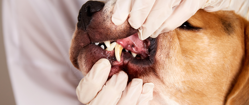
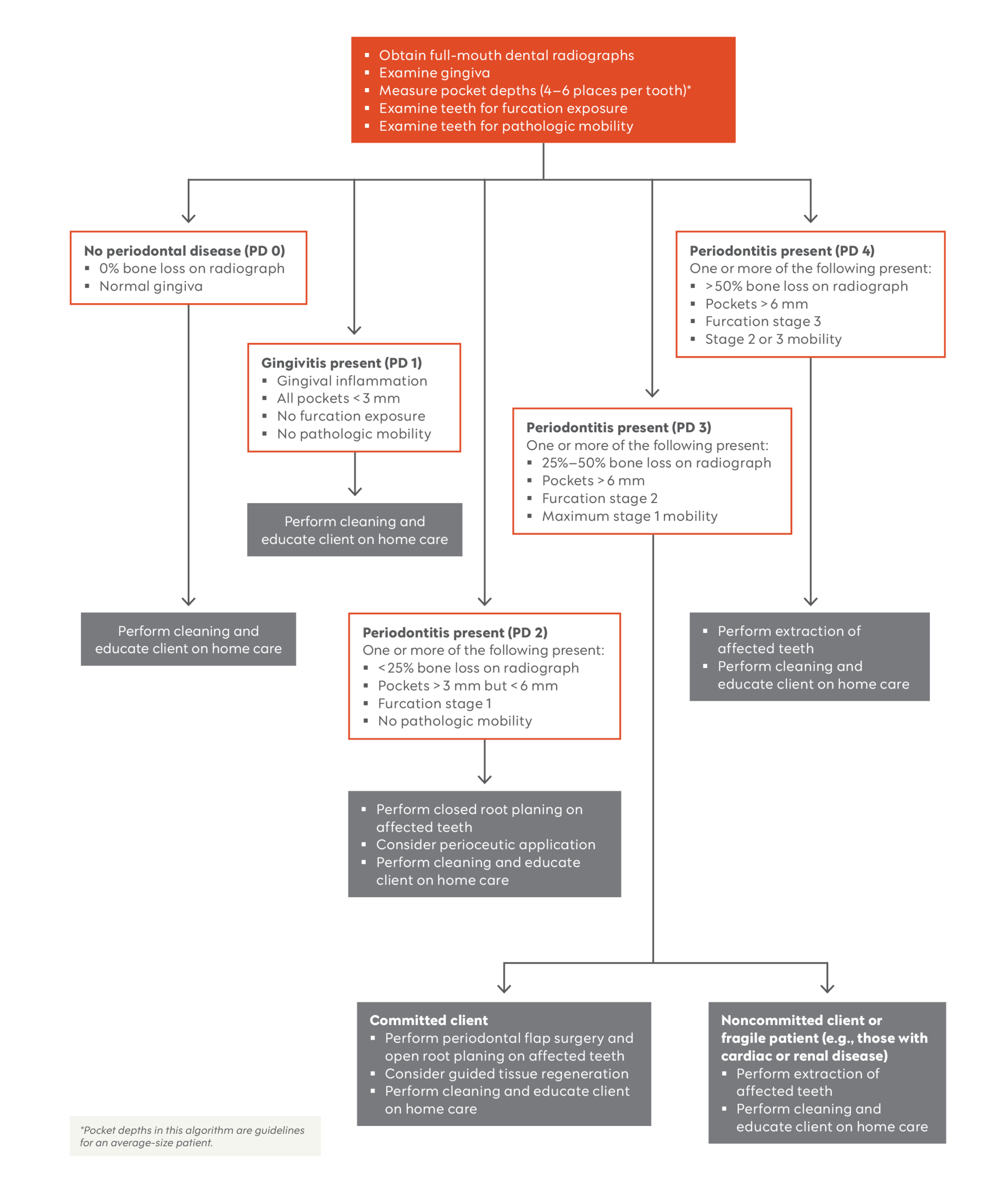
Proper decision making for therapy of periodontally diseased teeth can be challenging. Clinical factors that help determine the best therapy for a particular tooth include pocket depth in millimeters, percentage of bone attachment loss, furcation exposure (Box 1), and mobility (Box 2). These factors must be determined via periodontal probing and diagnostic imaging (intraoral dental radiology with or without cone beam computed tomography) performed while the patient is under anesthesia.
- Stage 1 (F1): Less than halfway under the crown in any direction of a multirooted tooth with attachment loss
- Stage 2 (F2): More than halfway under the crown of a multirooted tooth with attachment loss but not through and through
- Stage 3 (F3): Under the crown of a multirooted tooth, through and through from 1 side of the furcation and out the other
- Stage 0 (M0): Physiologic mobility up to 0.2 mm
- Stage 1 (M1): Mobility is increased in any direction other than axial over a distance between 0.2 mm and 0.5 mm
- Stage 2 (M2): Mobility is increased in any direction other than axial over a distance between 0.5 mm and 1 mm
- Stage 3 (M3): Mobility is increased in any direction other than axial over a distance greater than 1 mm or any axial movement
The whole clinical picture must be evaluated for each tooth. For example, healthy mandibular incisors often have apparent bone loss and mobility despite normal gingival attachment (< 3 mm sulcal depth in dogs).2-4
When to Perform Flap Surgery
Pocket depth and furcation involvement dictate the need to perform advanced periodontal flap surgery (Figure 1). Furcation 1 teeth can be cleaned without a flap (closed); however, cleaning furcation 2 (F2) and 3 (F3) teeth requires direct root visualization (flap).5,6 Periodontal flap surgery is also required when treating pockets deeper than 6 mm.4-10 If periodontal flap surgery is not performed in these cases, subgingival plaque, calculus, and granulation tissue will persist and the tooth will continue to have progressive disease until the next cleaning, as clients cannot effectively clean the hidden root surfaces at home.
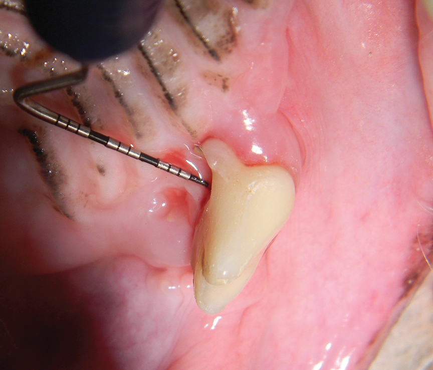
Figure 1. Maxillary fourth premolar with stage 2 furcation. This tooth cannot be effectively cleaned without a periodontal flap but has a very good prognosis with periodontal flap surgery and guided tissue regeneration. If referral is not an option, extraction is recommended.
When to Perform Extraction
Some teeth, particularly F3 teeth, cannot be preserved even with flap surgery and excellent home care. Based on the focal infection theory, periodontal inflammation creates both local and systemic effects.11,12 Thus, even if an F3 tooth with mild bone loss appears otherwise healthy and is cleaned with advanced therapies, periodontal inflammation will persist and progress with time (Figure 2). Likewise, F3 teeth with horizontal bone loss cannot be reliably returned to normal periodontal health, even with guided tissue repair.13-15 Bony repair can only happen in infrabony pockets and F2 teeth because bone grows laterally, not vertically. In the author’s opinion, all F3 teeth with bone loss should be extracted. Patients with severe disease and safety concerns or owners who wish to pursue preservation should be referred to a board-certified veterinary dentist.
Additional factors must also be considered. For example, is the client willing to perform home care? Does the patient have comorbidities that contraindicate future anesthesia? If home care will not be performed or the patient is an unsuitable candidate for additional anesthetic procedures, extraction should be recommended.
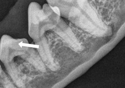
Figure 2. A premolar (307) with stage 3 furcation (through and through; arrow). This tooth should be extracted regardless of the fact that it has minimal bone loss.
Preventive Cleaning and Maintenance
Regardless of outward gingival appearance, most patients should have their teeth cleaned annually.16 Smaller-breed dogs or dogs and cats with increased plaque sensitivity that are more susceptible to periodontal disease, or animals that have received advanced periodontal flap procedures, may need anesthetized professional dental cleanings more frequently.
References
- AVDC nomenclature. American Veterinary Dental College. Accessed August 13, 2026. https://avdc.org/avdc-nomenclature
- Niemiec BA. Professional teeth cleaning. J Vet Dent. 2003;20(3):175-180.
- Niemiec B, Gawor J, Nemec A, et al. World Small Animal Veterinary Association Global Dental Guidelines. J Small Anim Pract. 2020;61(7):E36-E161. doi:10.1111/jsap.13113
- Stepaniuk K. Periodontology. In: Lobprise HB, Dodd JR, eds. Wiggs’s Veterinary Dentistry. 2nd ed. Wiley-Blackwell; 2019:81-108. doi:10.1002/9781118816219
- Theuns P. Furcation involvement and treatment. In: Niemiec BA, ed. Veterinary Periodontology. Wiley-Blackwell; 2013:289-295. doi:10.1002/9781118705018
- Huynh-Ba G, Kuonen P, Hofer D, Schmid J, Lang NP, Salvi GE. The effect of periodontal therapy on the survival rate and incidence of complications of multirooted teeth with furcation involvement after an observation period of at least 5 years: a systematic review. J Clin Periodontol. 2009;36(2):164-176. doi:10.1111/j.1600-051X.2008.01358.x
- Cobb CM. Non-surgical pocket therapy: mechanical. Ann Periodontol. 1996;1(1):443-490. doi:10.1902/annals.1996.1.1.443
- Niemiec BA. Advanced non-surgical therapy. In: Niemiec BA, ed. Veterinary Periodontology. Wiley-Blackwell; 2013:154-169. doi:10.1002/9781118705018
- Sanz M, Herrera D, Kebschull M, et al. Treatment of stage I-III periodontitis: the EFP S3 level clinical practice guideline. J Clin Periodontol. 2020;47 Suppl 22:4-60. doi:10.1111/jcpe.13290
- Zetner K, Rothmueller G. Treatment of periodontal pockets with doxycycline in beagles. Vet Ther. 2002;3(4):441-452.
- Niemiec BA. Local and regional consequences of periodontal disease. In: Niemiec BA, ed. Veterinary Periodontology. Wiley-Blackwell; 2013:69-80. doi:10.1002/9781118705018
- Niemiec BA. Systemic manifestations of periodontal disease. In: Niemiec BA, ed. Veterinary Periodontology. Wiley-Blackwell; 2013:81-90. doi:10.1002/9781118705018
- Lobprise HB, Stepaniuk K. Oral surgery – periodontal surgery. In: Lobprise HB, Dodd JR, eds. Wiggs’s Veterinary Dentistry. 2nd ed. Wiley-Blackwell; 2019:193-228. doi:10.1002/9781118816219
- Bezerra B. Pocket reduction therapy—resective approach. In: Newman MG, Klokkevold PR, Elangovan S, Kapila Y, eds. Newman and Carranza’s Clinical Periodontology. 14th ed. Elsevier;
2024:756-765. - Froum SJ, Weinberg MA, Rosenberg E, Tarnow D. A comparative study utilizing open flap debridement with and without enamel matrix derivative in the treatment of periodontal intrabony defects: a 12-month re-entry study. J Periodontol. 2001;72(1):25-34. doi:10.1902/jop.2001.72.1.25
- Niemiec BA. The complete dental cleaning. In: Niemiec BA, ed. Veterinary Periodontology. Wiley-Blackwell; 2013:129-153. doi:10.1002/9781118705018