
Respiratory infections in dogs may result from a wide range of infectious agents. Although parasites account for a small proportion of canine respiratory infections, they are often underdiagnosed when appropriate diagnostic testing is not performed. This article reviews the biology, clinical features, diagnosis, and treatment of parasitic respiratory infections in dogs. Practical diagnostic recommendations, including centrifugal fecal flotation; imaging; and endoscopy, when indicated, are outlined for evaluating dogs with respiratory signs. Applying a structured diagnostic approach will help clinicians distinguish parasitic from nonparasitic causes, leading to accurate treatment decisions and improved patient outcomes.
Take-Home Points
- In coughing dogs, the differential diagnosis should include heartworms and lungworms in endemic areas, as well as bacterial, viral, and noninfectious causes.
- Lungworms are less prevalent than other causes of respiratory infections overall; however, among lungworms, the prevalence of those that produce eggs as diagnostic stages is higher than that of those that produce larvae.
- In regions where heartworms are hyperendemic, heartworm testing is the first diagnostic step, followed by targeted fecal diagnostic tests.
- Using a combination of centrifugal fecal flotation with a high–specific-gravity solution and a Baermann test maximizes the likelihood of lungworm detection.
- Treatment with anthelmintics at the proper dose for an appropriate duration should follow specific diagnosis of lungworms.
Several infectious etiologic agents, including bacteria, viruses, fungi, and parasites, are responsible for upper and lower respiratory infections commonly encountered in veterinary practices.1 Since parasitic causes are rare, they may sometimes be misdiagnosed.2 However, when appropriate diagnostic tests are performed, parasitic causes can be ruled in or out with some confidence.
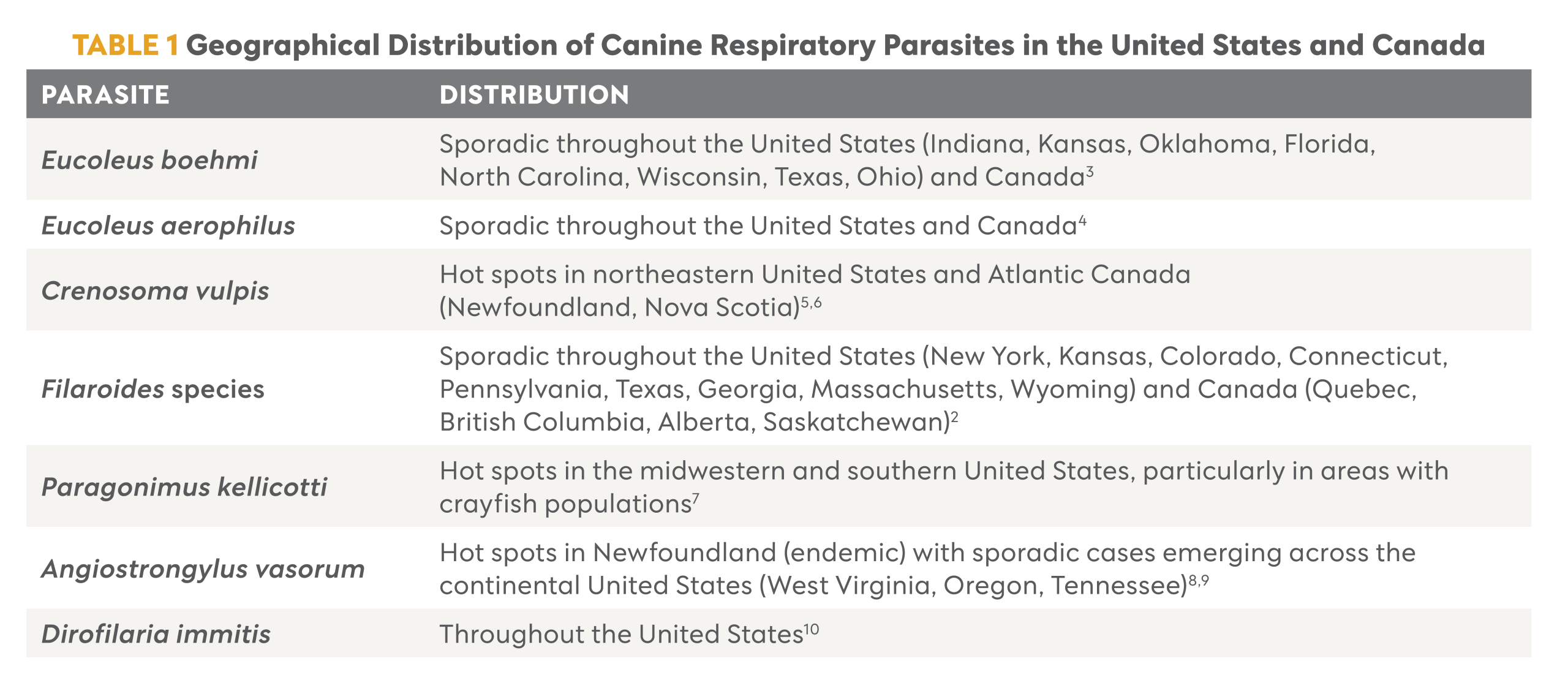
Lungworms in U.S. dogs include nematode parasites that live in the respiratory tract (Eucoleus aerophilus, Eucoleus boehmi, Crenosoma vulpis, Filaroides osleri, Filaroides hirthi, and Filaroides milksi) and a trematode parasite (Paragonimus kellicotti). Cardiopulmonary signs caused by Dirofilaria immitis must be differentiated from those associated with lungworm infections. In parts of eastern Canada, Angiostrongylus vasorum must also be considered. Distribution and hot spots of each species discussed in this article are listed in Table 1.
Eucoleus boehmi
E boehmi (previously known as Capillaria boehmi) adults live in the mucosal epithelium of the nasal passages and frontal and paranasal sinuses. Infection is acquired when eggs containing third-stage (L3) larvae (infective stages) are ingested from contaminated environments. Prevalence in U.S. dogs is 0.3%.11 Infections result in sneezing, mucopurulent nasal discharge, rhinitis, and epistaxis12; however, they can also be asymptomatic.
Female E boehmi worms produce bipolar, asymmetrical, golden-brown eggs (size, 54 to 60 × 30 to 35 µm) with fine pits on the surface (Figure 1A).13 Eggs are swallowed, pass into feces, and can be demonstrated by centrifugal fecal flotation methods. Rhinoscopic examination can demonstrate adult worms in situ.14,15
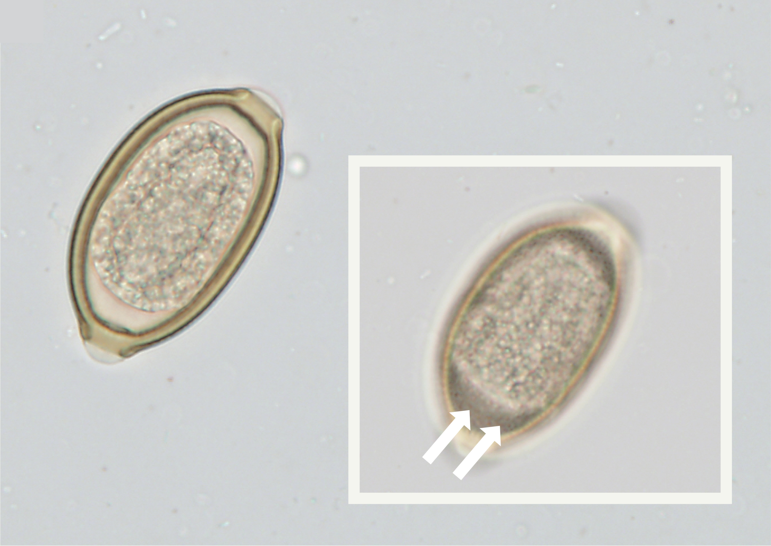
Figure 1A. Eggs of egg-producing lungworms. Eucoleus boehmi with inset showing the pitted surface of the egg (arrows).
There are no FDA-labeled treatments for E boehmi infection. Variable efficacy is reported with extralabel treatments. One dose of moxidectin 2.5 mg/kg topically was shown to be efficacious at reducing egg shedding by > 99%.15 Studies of milbemycin oxime and fenbendazole have shown variable efficacy. A single oral dose of milbemycin oxime 2 mg/kg was efficacious at eliminating egg shedding in 1 study16 but was ineffective at the same dose in another case.17 Similarly, fenbendazole at 50 mg/kg PO q24h for 10 to 14 days was successful in 2 studies,14,18 but a dosage of 100 mg/kg PO q24h for 2 weeks was ineffective in another report.17
Eucoleus aerophilus
E aerophilus (previously known as Capillaria aerophila) adults are slender worms that live in the trachea, bronchi, and bronchioles. Infection is acquired when eggs containing L3 larvae (infective stages) are ingested from contaminated environments. Sometimes, infective eggs are ingested by earthworms and dogs become infected by ingesting the earthworms. Prevalence in U.S. dogs is low at approximately 0.04%.11
Damage caused by worms results in chronic bronchitis, which may manifest clinically as chronic dry or moist cough, wheezing, sneezing, dyspnea, and general distress.19 Some infections are asymptomatic. Secondary bacterial infections may lead to complications such as bronchopneumonia and respiratory failure.4
Female E aerophilus worms lay bipolar, asymmetrical, brownish-green eggs (size, 58 to 79 × 29 to 40 µm) with a network of anastomosing ridges (Figure 1B).13,20 These are coughed up and swallowed and appear in feces and must be differentiated from Trichuris vulpis eggs, which are larger (size, 72 to 90 × 32 to 40 µm). Eggs can be demonstrated by centrifugal fecal flotation. Worms can be visualized by bronchoscopy, and eggs can be found in bronchoalveolar lavage samples.
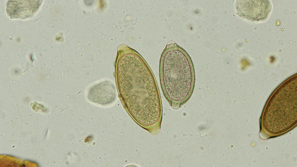
Figure 1B. Eggs of egg-producing lungworms. Eucoleus aerophilus egg (right) next to a Trichuris vulpis egg (left).
There are currently no FDA-labeled treatments for E aerophilus infection. Extralabel fenbendazole at 50 mg/kg PO q24h for 10 days and milbemycin oxime at 0.5 to 1.07 mg/kg PO given once have been used with success.21
Crenosoma vulpis
C vulpis (commonly known as fox lungworm) adults are small worms that live in the trachea, bronchi, and bronchioles. Wild carnivores—foxes and coyotes—serve as reservoir hosts. The name describes the annulation (crenations) on the outermost part (cuticle) of the worm body. Infection is acquired when the dog ingests slugs or snails containing the third larval stage.
C vulpis is restricted to the northeastern United States and Atlantic Canada, and prevalence in dogs is low in the United States (approximately 0.02%).11 In small regional pockets such as Prince Edward Island, C vulpis was responsible for 20.8% of coughing dogs in 1 study.22
Adult worms cause chronic bronchitis-bronchiolitis, resulting in infected animals experiencing coughing, gagging, and expectoration.6 Steroid injections may ameliorate the coughing temporarily.6 Infection also induces a mild eosinophilic inflammatory response.23
Female worms lay first-stage (L1) larvae, which are coughed up and swallowed and appear in feces. While the L1 larvae can be recovered by fecal flotation, the best method for confirmatory diagnosis is using a Baermann test on feces. Recovered larvae can be identified by length (264 to 340 × 16 to 22 µm) and tail morphology (straight, pointed tail with no indentations).13,24
A single oral dose of milbemycin oxime at 0.5 mg/kg was 98.7% effective at achieving clinical cure and stopped the shedding of L1 larvae in 2 studies.22,25 Fenbendazole at 50 mg/kg PO q24h for 3 days has also resulted in complete resolution.23
Filaroides species
F osleri (previously known as Oslerus osleri) adults reside in granulomatous nodules at the tracheal bifurcation, distal trachea, and bronchi. Infection is acquired by ingestion of the first larval stage in feces, vomit, or sputum. Prevalence of F osleri is extremely low in dogs, with 26 cases reported across 9 U.S. states in approximately 95 years.2 F osleri is more common in wildlife reservoirs such as coyotes and wolves.
Infection results in tracheobronchitis. Clinical signs range from acute dyspnea with sporadic, nonproductive cough to chronic cough; rarely, infection is asymptomatic. Adult worms produce eggs that quickly hatch to release L1 larvae, which are coughed up and swallowed and appear in feces. The L1 larvae do not migrate out of feces easily and are best demonstrated in a zinc sulfate fecal flotation rather than a Baermann test.26 Recovered larvae can be morphologically identified by length (232 to 266 µm) and an S-shaped tail with a deep dorsal indentation.13,24 Treatment with fenbendazole at 50 mg/kg PO q24h for 10 days or more has resulted in clinical resolution and recession of nodules.2
F hirthi and F milksi are sporadically found in dogs, although at a lower incidence than F osleri.27-29 Adults reside in the lung alveoli, parenchyma, and terminal bronchioles. Infections can result in pneumonia; however, most are subclinical. L1 larvae can be recovered by zinc sulfate fecal flotation. Treatment with fenbendazole at 50 mg/kg PO for 2 weeks with 3 high doses of ivermectin at 0.4 mg/kg SC every 2 weeks following diagnosis has resulted in cessation of larval shedding.30 Caution must be used in dogs with MDR1 (multidrug resistance 1 ) mutation when high doses of macrocyclic lactones are prescribed.
Paragonimus kellicotti
P kellicotti (commonly known as lung fluke) adults reside, typically as pairs, in cysts within the lung parenchyma. Infection is acquired by ingestion of uncooked crayfish and crabs that harbor the metacercarial stage. Prevalence in dogs is sporadic at 0.02%.11 Infection results in chronic coughing; wheezing; hemoptysis; sneezing; dyspnea; eosinophilia; and, rarely, pneumothorax.31,32 The cysts communicate with the bronchioles, allowing eggs produced by the flukes to be coughed up and swallowed and pass into feces. The eggs are large (75 to 118 × 42 to 67 µm) and golden brown, with a single operculum and distinctive shoulders (Figure 1C).13 Since the eggs are heavy, fecal sedimentation tests have higher sensitivity than fecal flotation.

Figure 1C. Eggs of egg-producing lungworms. Paragonimus kellicotti recovered in centrifugal fecal flotation using a high-specific-gravity flotation solution.
Treatment with fenbendazole at 50 mg/kg PO q24h for 14 days has resulted in cessation of egg shedding after 3 to 8 days of treatment, death of adult flukes, and resolution of clinical signs.33,34 Praziquantel at 23 to 25 mg/kg PO q8h for 2 to 3 days has also resulted in complete clinical resolution.35,36
Angiostrongylus vasorum
A vasorum (commonly known as French heartworm) adults reside in the pulmonary arteries and right heart. Infection is acquired when a dog ingests slugs or snails containing the third larval stage. Previously, A vasorum infections in North America were restricted to dogs and wild canids in Newfoundland, Canada. However, autochthonous cases have been reported from a dog in Oregon, a coyote in Tennessee, and a fox in West Virginia.8,37,38 Infection results in vascular damage in the pulmonary arteries and body responses to embolized eggs and larvae. Clinical signs include chronic coughing, dyspnea, exercise intolerance, anorexia, gagging, weight loss, secondary coagulation disorders, and sudden death.
L1 larvae are passed into feces and can be recovered by a Baermann test. Larvae can be distinguished by length (340 to 399 × 13 to 17 µm), presence of a dorsal notch, and a ventral indentation.13,24 An enzyme-linked immunosorbent assay for antigen detection is available in endemic regions.39
Treatment has been achieved with milbemycin oxime at 0.5 mg/kg PO every 7 days for 4 weeks.22 Moxidectin 2.5 mg/kg topically had 85.2% efficacy with a single application, while fenbendazole at 25 mg/kg PO q24h for 20 days had 91.3% efficacy.39
Diagnostic Testing
Fecal Tests
Fecal diagnostic tests offer a straightforward, inexpensive approach with high diagnostic yield in dogs with respiratory signs. A stepwise approach to diagnosing respiratory parasites in dogs is provided in Figure 2. In heartworm-hyperendemic areas such as the southern United States, veterinarians must test for D immitis antigen and microfilariae as recommended by the American Heartworm Society to rule out heartworms.40 A rapid, in-clinic blood/serum test to detect A vasorum antigen (IDEXX Angio Detect) is available in Canada and Europe. To detect the other lungworms described above, fecal flotation along with a concurrent Baermann test is recommended. If cost is an issue, begin with fecal flotation and follow with a Baermann test only if larvae are found.

Figure 2. A stepwise approach to diagnosing respiratory parasites in dogs.
Centrifugal fecal flotation using a high specific gravity solution is recommended as a first-line test in dogs of any age with respiratory signs in the United States as egg-producing parasites (Eucoleus and Paragonimus) are more prevalent than larvae-producing species (Filaroides and Crenosoma). These tests can be performed in-clinic or at diagnostic laboratories and recover both eggs and larvae. To improve diagnostic sensitivity, 2 or 3 fecal samples from consecutive days must be examined.26 Passive fecal flotation does not recover as many parasites as centrifugal flotation, resulting in missed diagnoses; therefore, passive flotation is not recommended.
If larvae are recovered on flotation using a high specific gravity solution, osmotic damage is likely. Zinc sulfate flotation solution may cause less damage to lungworm larvae.26 To confirm morphologic features of the larvae and identify the species, a Baermann test must be performed on fresh, nonrefrigerated feces. Feces must be collected immediately upon defecation, minimizing soil contamination, to avoid the entry of soil/plant/free-living nematodes into the sample. Since larval identification and differentiation from free-living larvae often require expertise, it is recommended that the Baermann test be performed at a state/commercial diagnostic laboratory along with a fecal flotation test even if flotation was performed in-clinic before. If clinical suspicion remains high despite a negative flotation result, a Baermann test is essential to rule out lungworm infection.
Imaging and Other Tests
Appropriate imaging modalities may help stage the patient and arrive at a diagnosis. Radiographic signs of parasitic infections have been reviewed by Birckhead et al.41 In E boehmi infections of the sinuses, no significant radiographic changes are seen. Since E aerophilus are an infection of the lower airways, radiographic changes are nonspecific and, depending on severity, can be a diffuse bronchial pattern, mixed bronchial and interstitial pattern, or alveolar pattern. In C vulpis infections, radiographic changes are nonspecific with a bronchial pattern and a concurrent alveolar pattern. Due to the location of F osleri at the tracheal bifurcation, radiographs may reveal intraluminal soft tissue opacities. These nodules can be confirmed by endoscopy. The lungs may have nonspecific bronchial patterns. In F hirthi infections, lungs show patchy to diffuse interstitial to alveolar patterns. In P kellicotti infections, radiographs reveal focal patchy infiltrates, pulmonary cavitations and pneumothorax, whereas on computed tomography images, lesions appear as hypodense nodules with rim enhancement.31
Endoscopic modalities include rhinoscopy for examining the nasal passages in E boehmi infections to visualize and recover adult nematodes and eggs.14 Bronchoscopy can be used to examine the lower airways for nodules in F osleri infections and adult worms in E aerophilus, C vulpis, and other infections. Transtracheal/bronchoalveolar washes may have elevated eosinophils and may contain parasite eggs/larvae. In most lungworm infections, peripheral eosinophilia can be a supportive finding; however, its absence does not rule out lungworm infection.
Summary
In clinical practice, fecal diagnostics provide a simple and affordable way to screen dogs with respiratory signs for parasitic infections. Starting with centrifugal fecal flotation and adding a Baermann test when larvae are suspected offers the best balance between accuracy and cost. Pairing these results with appropriate imaging and/or endoscopy helps confirm the diagnosis, determine the extent of disease, and guide targeted and adjunct treatments. Using a systematic approach ensures that parasitic causes of respiratory disease are not overlooked and patients receive timely care.
References
- Vieson MD, Piñeyro P, LeRoith T. A review of the pathology and treatment of canine respiratory infections. Vet Med (Auckl). 2012;3:25-39. doi:10.2147/VMRR.S25021
- Yao C, O’Toole D, Driscoll M, et al. Filaroides osleri (Oslerus osleri): two case reports and a review of canid infections in North America. Vet Parasitol. 2011;179(1-3):123-129. doi:10.1016/j.vetpar.2011.02.012
- Gillis-Germitsch N, Müller S, Gori F, et al. Capillaria boehmi (syn. Eucoleus boehmi): challenging treatment of a rarely diagnosed nasal nematode in dogs and high prevalence in Swiss foxes. Vet Parasitol. 2020;281:109103. doi:10.1016/j.vetpar.2020.109103
- Samorek-Pieróg M, Cencek T, Łabuć E, et al. Occurrence of Eucoleus aerophilus in wild and domestic animals: a systematic review and meta-analysis. Parasit Vectors. 2023;16(1):245. doi:10.1186/s13071-023-05830-0
- Bihr T, Conboy GA. Lungworm (Crenosoma vulpis) infection in dogs on Prince Edward Island. Can Vet J. 1999;40(8):555-559.
- Pohly AG, Nijveldt EA, Stone MS, Walden HD, Ossiboff RJ, Conrado FO. Infection with the fox lungworm (Crenosoma vulpis) in two dogs from New England: two clinical reports and updated geographic distribution in North America. Vet Parasitol Reg Stud Reports. 2022;30:100714. doi:10.1016/j.vprsr.2022.100714
- Diaz JH. Paragonimiasis acquired in the United States: native and nonnative species. Clin Microbiol Rev. 2013;26(3):493-504. doi:10.1128/CMR.00103-12
- Riese K, Baker E, Dennis MM, Williamson R, Gerhold R. Two cases of Angiostrongylus vasorum, a cardiopulmonary nematode, in a wild black bear and coyote of Tennessee. Vet Parasitol Reg Stud Reports. 2024;54:101079. doi:10.1016/j.vprsr.2024.101079
- Conboy GA. Canine angiostrongylosis: the French heartworm: an emerging threat in North America. Vet Parasitol. 2011;176(4):382-389. doi:10.1016/j.vetpar.2011.01.025
- Drake J, Wiseman S. Increasing incidence of Dirofilaria immitis in dogs in USA with focus on the southeast region 2013-2016. Parasit Vectors. 2018;11(1):39. doi:10.1186/s13071-018-2631-0
- Sobotyk C, Upton KE, Lejeune M, et al. Retrospective study of canine endoparasites diagnosed by fecal flotation methods analyzed across veterinary parasitology diagnostic laboratories, United States, 2018. Parasit Vectors. 2021;14(1):439. doi:10.1186/s13071-021-04960-7
- Conboy G. Helminth parasites of the canine and feline respiratory tract. Vet Clin North Am Small Anim Pract. 2009;39(6):1109-1126. doi:10.1016/j.cvsm.2009.06.006
- Zajac AM, Conboy GA, Little SE, Reichard MV. Fecal examination for the diagnosis of parasitism. In: Zajac AM, Conboy GA, Little SE, Reichard MV, eds. Veterinary Clinical Parasitology. 9th ed. Wiley-Blackwell; 2021:1-190.
- Baan M, Kidder AC, Johnson SE, Sherding RG. Rhinoscopic diagnosis of Eucoleus boehmi infection in a dog. JAAHA. 2011;47(1):60-63. doi:10.5326/JAAHA-MS-5707
- Veronesi F, Di Cesare A, Braun G, et al. Evaluation of the clinical efficacy and safety of a spot-on combination of imidacloprid 10 % / moxidectin 2.5 % (Advocate®, Advantage® Multi) in comparison to an untreated control group in the treatment of Capillaria boehmi in naturally infected dogs. Parasitol Res. 2017;116(suppl 1):65-74. doi:10.1007/s00436-017-5492-0
- Conboy G, Stewart T, O’Brien S. Treatment of E. boehmi infection in a mixed-breed dog using milbemycin oxime. JAAHA. 2013;49(3):204-209. doi:10.5326/JAAHA-MS-5832
- Alho AM, Mouro S, Pissarra H, et al. First report of Eucoleus boehmi infection in a dog from Portugal. Parasitol Res. 2016;115(4):1721-1725. doi:10.1007/s00436-016-4932-6
- Burgess H, Ruotsalo K, Peregrine AS, Hanselman B, Abrams-Ogg A. Eucoleus aerophilus respiratory infection in a dog with Addison’s disease. Can Vet J. 2008;49(4):389-392.
- Traversa D, Di Cesare A, Milillo P, Iorio R, Otranto D. Infection by Eucoleus aerophilus in dogs and cats: is another extra-intestinal parasitic nematode of pets emerging in Italy? Res Vet Sci. 2009;87(2):270-272. doi:10.1016/j.rvsc.2009.02.006
- Traversa D, Di Cesare A, Lia RP, et al. New insights into morphological and biological features of Capillaria aerophila (Trichocephalida, Trichuridae). Parasitol Res. 2011;109(suppl 1):97-104. doi:10.1007/s00436-011-2406-4
- Abbate JM, Napoli E, Arfuso F, et al. Six-month field efficacy and safety of the combined treatment of dogs with Frontline Tri-Act® and NexGard Spectra®. Parasit Vectors. 2018;11(1):425. doi:10.1186/s13071-018-2957-7
- Conboy G. Natural infections of Crenosoma vulpis and Angiostrongylus vasorum in dogs in Atlantic Canada and their treatment with milbemycin oxime. Vet Rec. 2004;155(1):16-18. doi:10.1136/vr.155.1.16
- Peterson EN, Barr SC, Gould W 3rd, Beck KA, Bowman DD. Use of fenbendazole for treatment of Crenosoma vulpis infection in a dog. JAVMA. 1993;202(9):1483-1484.
- McGarry J, Morgan E. Identification of first-stage larvae of metastrongyles from dogs. Vet Rec. 2009;165(9):258-261. doi:10.1136/vr.165.9.258
- Conboy G, Bourque A, Miller L, Seewald W, Schenker R. Efficacy of Milbemax (milbemycin oxime+ praziquantel) in the treatment of dogs experimentally infected with Crenosoma vulpis. Vet Parasitol. 2013;198(3-4):319-324. doi:10.1016/j.vetpar.2013.09.016
- Traversa D, Di Cesare A, Conboy G. Canine and feline cardiopulmonary parasitic nematodes in Europe: emerging
and underestimated. Parasit Vectors. 2010;3:62. doi:10.1186/1756-3305-3-62 - Georgi JR, Fahnestock GR, Bohm MF, Adsit JC. The migration and development of Filaroides hirthi larvae in dogs. Parasitology. 1979;79(1):39-47. doi:10.1017/s0031182000051969
- Georgi JR, Georgi ME, Cleveland DJ. Patency and transmission of Filaroides hirthi infection. Parasitology. 1977;75(2):251-257. doi:10.1017/s0031182000062399
- Greenway JA, Stockdale PH. Case report. A case tentatively diagnosed as Filaroides milksi in a dog. Can Vet J. 1970;11(10):203-204.
- Cervone M, Giannelli A, Rosenberg D, Perrucci S, Otranto D. Filaroidosis infection in an immunocompetent adult dog from France. Helminthologia. 2018;55(1):77-83. doi:10.1515/helm-2017-0058
- Charlebois PR, Bersenas AM, Yiew XT, Weese JS, Peregrine AS. Local outbreak of spontaneous pneumothorax secondary to paragonimosis in southwestern Ontario dogs. Can Vet J. 2023;64(7):643-649.
- Dubey JP. Endemic Paragonimus kellicotti infections in animals and humans in USA and Canada: review and personal perspective. Food Waterborne Parasitol. 2023;30:e00184. doi:10.1016/j.fawpar.2022.e00184
- Dubey JP, Miller TB, Sharma SP. Fenbendazole for treatment of Paragonimus kellicotti infection in dogs. JAVMA. 1979;174(8):835-837.
- Saini N, Ranjan R, Singla LD, Anand A, Randhawa CS. Successful treatment of pulmonary paragonimiasis in a German shepherd dog with fenbendazole. J Parasit Dis. 2012;36(2):171-174. doi:10.1007/s12639-012-0098-z
- Bowman DD, Frongillo MK, Johnson RC, Beck KA, Hornbuckle WE, Blue JT. Evaluation of praziquantel for treatment of experimentally induced paragonimiasis in dogs and cats. Am J Vet Res. 1991;52(1):68-71.
- Kirkpatrick CE, Shelly EA. Paragonimiasis in a dog: treatment with praziquantel. JAVMA. 1985;187(1):75-76.
- Williams LBA, Buswell ML, Perisho NA. First autochthonous case of Angiostrongylus vasorum in a domestic dog in the United States. JAVMA. 2024;262(10):1-3. doi:10.2460/javma.24.06.0408
- Kistler WM, Brown JD, Allison AB, Nemeth NM, Yabsley MJ. First report of Angiostrongylus vasorum and Hepatozoon from a red fox (Vulpes vulpes) from West Virginia, USA. Vet Parasitol. 2014;200(1-2):216-220. doi:10.1016/j.vetpar.2013.12.007
- Di Cesare A, Traversa D. Canine angiostrongylosis: recent advances in diagnosis, prevention, and treatment. Vet Med (Auckl). 2014;5:181-192. doi:10.2147/VMRR.S53641
- American Heartworm Society. American Heartworm Society canine guidelines for the prevention, diagnosis, and management of heartworm (Dirofilaria immitis) infection in dogs. Revised 2024. Accessed May 6, 2026. https://www.heartwormsociety.org/veterinary-resources/american-heartworm-society-guidelines
- Birckhead A, Malik R, Shamsi S, Jenkins D, Carstens A. A review of radiographic and computed tomographic features of helminth and arthropod parasitic diseases in dogs. Vet Radiol Ultrasound. 2025;66(6):e70108. doi:10.1111/vru.70108