
To take the CE quiz, click here.
This quiz is open until June 2028.

Deep fungal dermatoses in cats are uncommon but potentially serious infections. They may be confined to the skin or may originate from, or disseminate to, other locations, such as the respiratory tract or central nervous system. Key diseases include sporotrichosis, cryptococcosis, histoplasmosis, blastomycosis, coccidioidomycosis, phaeohyphomycosis, and pythiosis. Clinical signs vary widely and frequently resemble bacterial infection, immune-mediated disease, or neoplasia. Consideration of geographic location, environmental exposure, and travel history is essential when formulating differential diagnoses. Definitive diagnosis typically requires integration of cytology, histopathology with special stains, serologic testing, diagnostic imaging, and fungal culture with molecular confirmation. Prompt diagnosis and appropriate antifungal therapy are critical to optimize clinical outcomes and inform prognosis.
Take-Home Points
- Apply a structured, stepwise approach to the evaluation of suspected deep fungal dermatoses in cats by integrating signalment, exposure history, cytology, advanced diagnostics, and appropriate therapeutic planning.
- Recognize key morphologic features of major feline deep fungal pathogens to guide provisional diagnosis and appropriate confirmatory testing.
- Select appropriate first-line and adjunctive antifungal therapies based on pathogen identification, disease distribution, and organ involvement.
Deep fungal dermatoses in cats are uncommon but clinically significant and may result in substantial morbidity or even death. Cats typically present with firm to fluctuant dermal or subcutaneous nodules or ill-defined swellings that may ulcerate and form draining tracts. Lesions may be solitary or multifocal and can exude serosanguineous to purulent material. Importantly, nodular or ulcerative skin disease does not necessarily indicate infection.
The most critical step is distinguishing infectious from noninfectious causes, as treatment and prognosis differ markedly. Misdiagnosing a fungal infection as an immune-mediated condition can be dangerous; immunosuppressive therapy, particularly glucocorticoids, may worsen local invasion and facilitate systemic spread. Therefore, an accurate diagnosis before initiating treatment is essential.
In North America, important pathogens include species of Sporothrix, Cryptococcus, Histoplasma, Blastomyces, Coccidioides, and melanized (dematiaceous) fungi. Pythium, although not a true fungus, can produce clinically similar invasive disease and is increasingly recognized. Infection typically follows inhalation of environmental spores or traumatic inoculation. Thus, a systematic diagnostic strategy, including careful history review, cytology, histopathology, fungal culture, molecular techniques, and antigen/antibody testing, facilitates accurate pathogen identification and supports timely clinical decision-making. Box 1 summarizes the red flags for suspicion of deep fungal dermatoses in cats, and Figure 1 illustrates the step-by-step clinical diagnostic algorithm for deep fungal dermatoses in cats.
- Soil, vegetation, bird/bat guano, water, trauma
2. Chronic, nonhealing ulcers, nodules, or draining tracts
- Unresponsive or partially responsive to antibacterial treatment
3. Rapid worsening after immunosuppressive therapy
- Reconsider infectious etiology immediately
4. Granulomatous/pyogranulomatous inflammation on cytology
- Other differentials include bacterial infections, immune-mediated disorders, and foreign body reactions
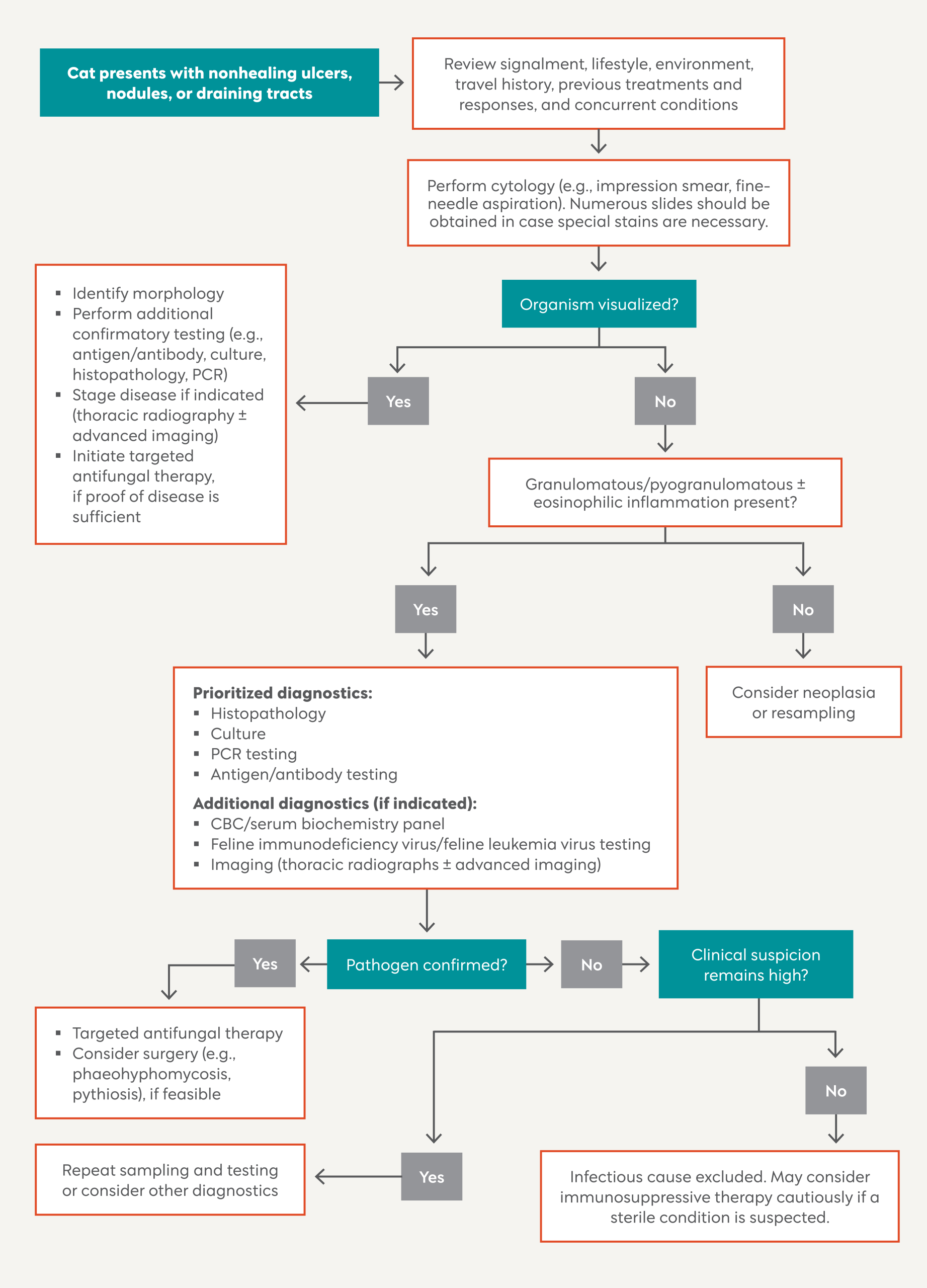
Figure 1. Diagnostic algorithm for deep fungal dermatoses in cats.
History and Physical Examination
Signalment
Deep fungal infections can affect cats regardless of age, breed, or sex; however, specific predispositions have been documented. For example, cryptococcosis is often seen in young adult cats, and sporotrichosis is most frequently identified in young, sexually intact, free-roaming male cats, likely due to increased environmental exposure and cutaneous inoculation from fighting and trauma.1 While many affected cats are immunocompetent, severe or disseminated disease may be more likely in immunocompromised
individuals.
Lifestyle, Environment, and Travel History
A thorough residence and travel history is essential, as many pathogens are associated with defined endemic regions, such as semiarid desert areas (e.g., Arizona) for coccidioidomycosis.2 Geographic location can substantially influence the likelihood of specific mycoses and should always be interpreted in the context of travel history.
Environmental exposure plays a central role in pathogenesis. Inhalation of environmental spores is the most common route of infection for most systemic fungi, whereas some fungal infections are typically acquired through traumatic inoculation of the skin. Outdoor access; hunting behavior; and exposure to soil, decaying vegetation, bird guano, or standing water should be carefully documented. Importantly, indoor-only cats are not exempt from risk. Many fungal spores are airborne or may be introduced into the home through contaminated organic materials, including potting soil. Therefore, absence of outdoor access does not eliminate the possibility of deep mycosis.
Previous Treatments and Responses
A typical history includes chronic, nonhealing nodules or draining tracts that showed limited to partial response to multiple courses of empirical antibiotics. Temporary improvement may reflect control of secondary bacterial infection rather than cure. A particular red flag is rapid progression or systemic spread after initiation of immunosuppressive medications (e.g., glucocorticoids, cyclosporine) prescribed for a presumed immune-mediated condition. Such deterioration should immediately prompt reconsideration of an infectious etiology.
Concurrent Conditions
Although some cats with deep fungal infections are systemically immunocompetent, concurrent diseases can affect susceptibility, treatment response, and prognosis. Retroviral testing for feline immunodeficiency virus (FIV) and feline leukemia virus (FeLV) is recommended, as infection may impair immune function and increase relapse risk. Neoplasia or prior immunosuppressive therapy should also be documented, as immunosuppression predisposes to dissemination and complicates management.
Determining the Cause
Assess Cytology
Cytology is a critical first-line diagnostic tool because it is rapid, is minimally invasive, and often yields clinically actionable information. Fine-needle aspiration of intact nodules or lymph nodes, along with impression smears from ulcerated areas or draining tracts, should be routinely performed and evaluated using Romanowsky-type stains (e.g., Diff-Quik). Identification of pyogranulomatous or granulomatous inflammation should raise suspicion for fungal infection.
Organisms may appear as yeasts (Sporothrix, Cryptococcus, Histoplasma, Blastomyces), pigmented hyphae (dematiaceous fungi), or negatively stained hyphae (Pythium). However, failure to visualize organisms does not exclude infection. If cytology reveals a pathogen consistent with the clinical presentation, subsequent diagnostics should focus on confirmation and speciation.
Choose Further Diagnostics
If cytology is inconclusive or suggests infection without definitive organism identification, additional diagnostics are necessary to confirm the pathogen, assess systemic involvement, and guide treatment planning.
CBC and Biochemistry
Systemic fungal infections frequently produce nonspecific laboratory abnormalities. Common findings include an inflammatory leukogram, mild to marked hyperglobulinemia, and nonregenerative anemia. Hypercalcemia may occur secondary to macrophage activation within granulomatous lesions. Serum biochemistry also establishes baseline hepatic and renal function prior to prolonged antifungal therapy. Concurrent testing for FIV/FeLV is recommended, as retroviral status may influence treatment duration, clinical response, and overall prognosis.
Fungal-Specific Tests
Antigen and antibody assays can provide valuable, minimally invasive diagnostic information and are particularly useful early in the workup. For example, the cryptococcal capsular polysaccharide antigen test (latex agglutination) demonstrates high sensitivity (95%) when performed on serum or cerebrospinal fluid.3 Urine antigen enzyme-linked immunosorbent assay (ELISA) for Histoplasma is similarly sensitive (90% to 94.4%),4,5 although cross-reactivity with Blastomyces may occur. For suspected coccidioidomycosis, antibody detection via agar gel immunodiffusion (AGID) remains the preferred diagnostic modality, with reported sensitivity of 97.7% to 100%.2,6
Diagnostic Imaging
Thoracic radiography is often recommended, as inhalation is the primary route of some infections. Advanced imaging (computed tomography or magnetic resonance imaging) may be used when evaluating sinonasal, orbital, or central nervous system disease and for determining the full extent of regional invasion.
Histopathology
Histopathology is commonly employed to characterize the pattern of cutaneous lesions, classify disease, exclude neoplasia, assess depth of invasion, and ideally identify and provisionally classify fungal organisms. Multiple deep biopsies are recommended, as fungal elements are often concentrated within necrotic centers of lesions. Routine hematoxylin and eosin staining may not always reliably highlight fungal structures; therefore, special stains such as periodic acid–Schiff and Grocott methenamine silver, among others, are sometimes necessary to enhance visualization.
Fungal Culture
Culture of fresh tissue biopsy specimens, rather than swabs, significantly improves the likelihood of isolating the causative fungal organism. Laboratories should be notified in advance if highly infectious and zoonotic pathogens such as Blastomyces, Histoplasma, or Coccidioides are suspected. Culture of these organisms should be performed only when clinically necessary.
Molecular Diagnostics
Panfungal PCR testing targeting the internal transcribed spacer regions can support species identification. It may be performed on cultured isolates and on fresh, frozen, or formalin-fixed paraffin-embedded (FFPE) tissues when culture is unavailable or negative. In one study, amplifiable fungal DNA was obtained from FFPE samples in 69.5% of cases, with 61.7% agreement between molecular and histologic results.7 PCR testing can also be applied to stained cytology slides, showing up to 86% sensitivity when fungal organisms are abundant.8 Since this technique may detect environmental contamination, results must be interpreted in conjunction with clinical presentation, cytology, and histopathology.
Determining the Treatment
If no pathogen is identified despite a thorough diagnostic investigation, noninfectious conditions such as sterile nodular panniculitis should be considered. These disorders often require immunosuppressive therapy, which must be administered cautiously and monitored closely. Rapid clinical deterioration following immunosuppression should prompt reconsideration of an infectious cause and repeat diagnostic testing.
If a specific pathogen is identified and consistent with the clinical presentation, antifungal therapy should be initiated and appropriate additional tests performed to assess the extent of disease. Itraconazole is commonly used as first-line medical therapy for sporotrichosis, histoplasmosis, and blastomycosis, whereas fluconazole is often preferred for cryptococcosis and coccidioidomycosis due to its superior penetration into the central nervous system when neurologic involvement is present. Posaconazole is a broader-spectrum triazole that may offer improved efficacy in some refractory cases, and pharmacokinetic data are available to support use of the oral suspension formulation.9 Amphotericin B may be indicated for severe, disseminated, or life-threatening infections; the deoxycholate formulation is effective but associated with greater nephrotoxicity, whereas lipid complex formulations are better tolerated but more costly.
In selected cases of phaeohyphomycosis and pythiosis, complete surgical excision may be the treatment of choice when feasible. Medical treatment is typically prolonged for several months and should continue beyond clinical resolution. Close monitoring for adverse effects and relapse is essential.
Selected Deep Fungal Dermatoses
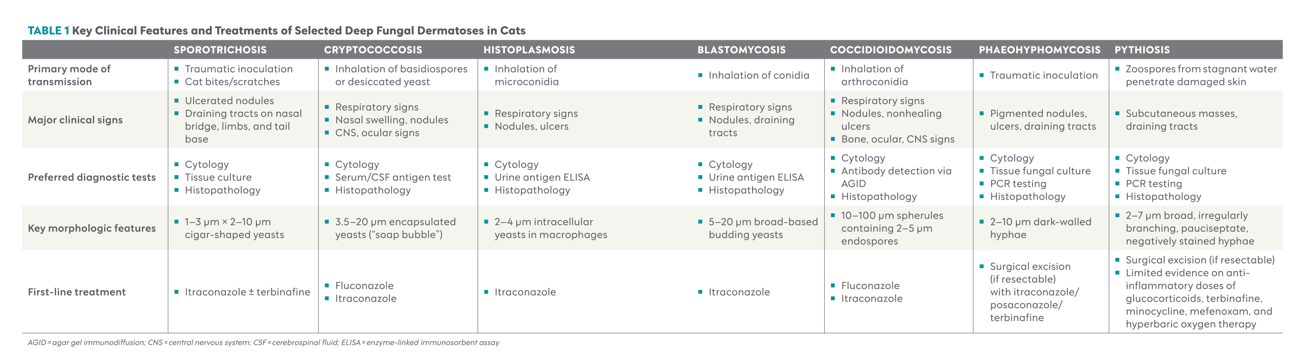
Key features and treatments of the following deep fungal dermatoses are summarized in Table 1.
Sporotrichosis
Sporotrichosis is caused by the dimorphic fungi Sporothrix schenckii species complex. Sporothrix brasiliensis has primarily been reported in South America, specifically Brazil and Chile, whereas S schenckii has a worldwide distribution. Infection follows traumatic inoculation with contaminated plant material; however, horizontal transmission through cat bites and scratches is also common, especially among free-roaming intact males. Cats often harbor large numbers of organisms and represent an important zoonotic source. Owners should wear gloves and protective clothing, and, in severe cases, respiratory and eye protection when handling affected cats.
Dermatologic lesions include multifocal ulcerated nodules and draining tracts, frequently involving the nasal bridge, distal limbs, or tail base. Cytology is often highly diagnostic in cats, revealing pleomorphic oval- to cigar-shaped yeasts measuring approximately 1 to 3 µm wide and 2 to 10 µm long, typically observed within macrophages,10 whereas deep tissue culture remains the most reliable diagnostic test (sensitivity exceeds 98%).1
Itraconazole is the preferred first-line therapy for S brasiliensis, whereas combination therapy with terbinafine and itraconazole may provide synergistic benefit in cases caused by S schenckii.11
Cryptococcosis
Cryptococcosis is most commonly caused by the dimorphic fungi Cryptococcus gattii or Cryptococcus neoformans, acquired primarily through inhalation of environmental basidiospores or desiccated yeast from soil, plant debris, or pigeon guano. C neoformans is distributed worldwide, whereas C gattii occurs in regions such as Western Europe, Australia, southern Africa, the Pacific Northwest, and coastal California.
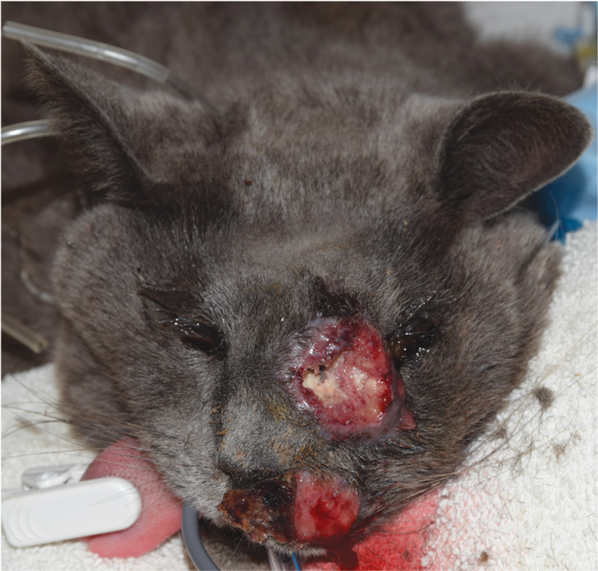
The nasal cavity is the usual site of initial infection, with potential extension to the skin, eyes, or central nervous system. Affected cats often present with nasal swelling (“Roman nose”), firm cutaneous nodules, ulceration, or neurologic signs (Figure 2). Cytology frequently reveals numerous 3.5- to 20-µm oval yeasts with a thick, clear mucopolysaccharide capsule (“soap bubble”) and narrow-based budding.12 Detection of cryptococcal capsular antigen in serum or cerebrospinal fluid is highly sensitive and specific and is the preferred diagnostic test.3
Fluconazole is commonly administered, especially in cats with neurologic involvement, although itraconazole may be considered in refractory cases.13

Histoplasmosis
Histoplasmosis is caused by the dimorphic fungus Histoplasma capsulatum. H capsulatum favors tropical and subtropical climates. Areas of intense endemicity include the Ohio and Mississippi River valleys, South America, and regions of southeast Asia and India. Infection most commonly results from inhalation of airborne microconidia in soil contaminated with bird or bat guano, and even indoor-only cats may be at risk.
Disease is often systemic, with clinical signs such as lethargy, respiratory distress, or lameness, and cutaneous nodules or ulcers are reported in 16.8% of cases.14 Urine antigen ELISA serves as a highly sensitive (90% to 94.4%) but less specific (64.6%) noninvasive screening test.4,5 However, a positive result is not definitively diagnostic as cross-reactivity with other fungi, including Blastomyces, can occur. Definitive diagnosis is based on identification of small (2 to 4 µm) yeasts with a clear halo within macrophages or neutrophils on cytology or histopathology.10 Fungal culture is generally avoided due to biohazard risk.
Itraconazole is the preferred first-line therapy.
Blastomycosis
Blastomycosis results primarily from the dimorphic fungi Blastomyces dermatitidis or Blastomyces gilchristii and, rarely, Blastomyces helicus. Infection occurs following inhalation of airborne conidia from moist, acidic soil, particularly in regions surrounding waterways. In North America, cases are most frequently reported in the Mississippi, Missouri, and Ohio River valleys and parts of the mid-Atlantic.
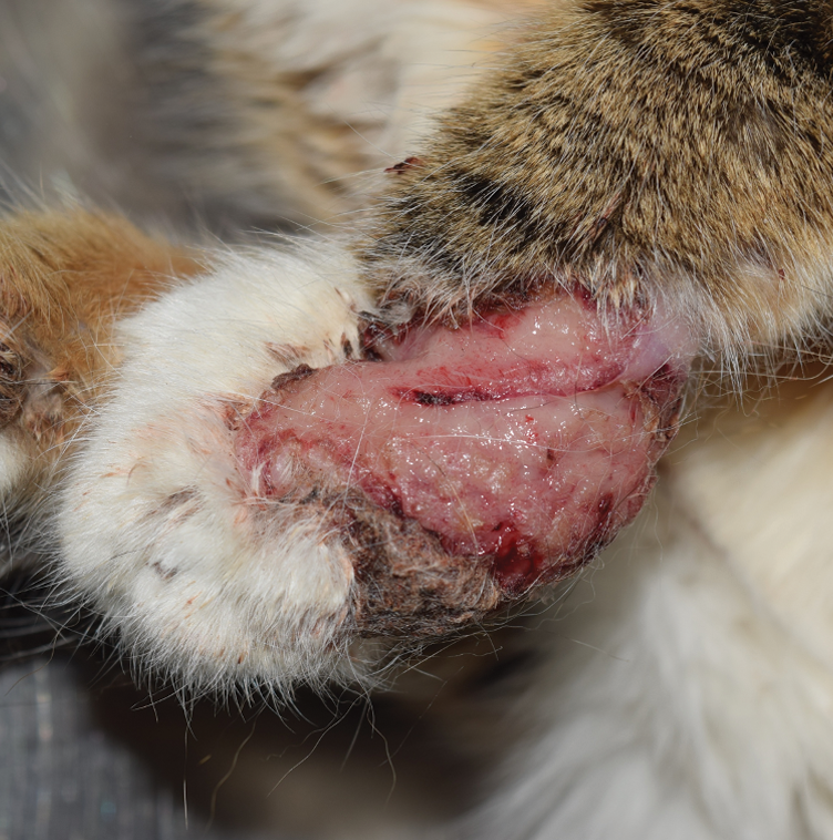
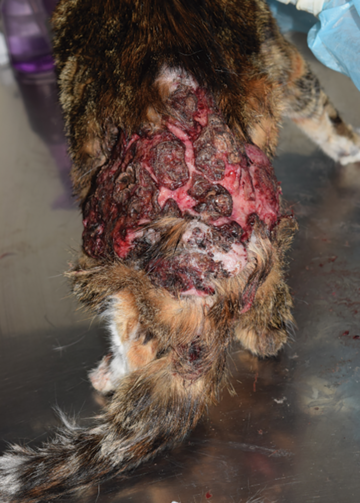
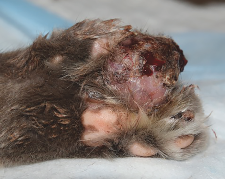
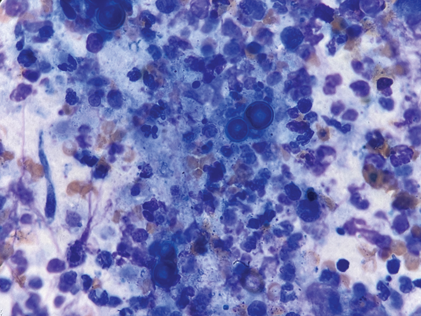
Affected cats commonly develop respiratory signs, including dyspnea and coughing, in addition to cutaneous nodules or draining tracts (Figure 3). Cytology and histopathology typically reveal large (5 to 20 µm),10 thick-walled yeasts with broad-based budding within pyogranulomatous inflammation (Figure 4). Urine antigen ELISA is highly sensitive (93.5% to 100%) but cross-reacts with Histoplasma, limiting specificity.15,16
Itraconazole is the preferred first-line therapy.
Coccidioidomycosis
Coccidioidomycosis is caused by the dimorphic fungi Coccidioides immitis and Coccidioides posadasii. Infection follows inhalation of airborne arthroconidia from semiarid, alkaline soils in endemic regions (Arizona and south-central valley of California); notably, many affected cats are strictly indoor.2
Clinical signs most commonly include respiratory manifestations such as cough, tachypnea, and dyspnea. With dissemination to the skin, cats may develop nodules, nonhealing ulcers, or plaque-like crusting, especially on the trunk and distal extremities. Fever, lameness due to bone involvement, and ocular or neurologic abnormalities may also occur.
Morphologically, large (10 to 100 µm), thick, double-walled spherules containing endospores (2 to 5 µm) are observed.10,12 However, since organisms are often sparse in samples, antibody detection using AGID is the primary diagnostic test (sensitivity 97.7% to 100%).2,6
Fluconazole is generally the preferred first-line therapy, although itraconazole may be favored in cases with bone involvement.
Phaeohyphomycosis
Phaeohyphomycosis is caused by dematiaceous, or melanized, fungi such as Alternaria, Exophiala, Curvularia, Fonsecaea, and neurotropic Cladophialophora (formerly Xylohypha or Cladosporium), among others. These organisms have a worldwide distribution. Infection typically follows traumatic cutaneous inoculation from contaminated soil or plant material.
Affected cats commonly develop solitary or multifocal nodules, ulcers, or draining tracts, often involving the nose, ears, or digits. Because of dark coloration and nodular appearance, these lesions can be misidentified as melanoma. Cytology and histopathology may identify dark-walled hyphae measuring 2 to 10 µm.12 Fungal culture and panfungal PCR testing aid in species identification.
Management generally consists of aggressive surgical excision, when feasible, combined with prolonged systemic antifungal therapy such as itraconazole, terbinafine, posaconazole, or combination regimens.
Pythiosis
Pythiosis is caused by the aquatic oomycete Pythium insidiosum. P insidiosum is widely distributed in tropical and subtropical regions, including the Gulf Coast and southeastern United States (particularly Florida), and has also been reported across the Americas, Europe, Asia, and Australia, reflecting global presence. Infection presumptively occurs when motile zoospores in warm, stagnant water penetrate damaged skin or gastrointestinal mucosa.
In cats, disease is primarily cutaneous and locally aggressive, presenting as subcutaneous masses and draining tracts, commonly affecting the inguinal and perianal regions, tail base, limbs, or neck. Cytology and histopathology typically reveal marked pyogranulomatous inflammation with eosinophilic sleeves around broad (2 to 7 µm), irregularly branching, pauciseptate, negatively stained hyphae.12 Definitive diagnosis relies on culture and/or PCR testing.
Treatment is challenging as oomycetes lack ergosterol, a target of many antifungals. Aggressive surgical excision with wide margins is recommended, often combined with antifungal therapies, although supporting evidence remains limited. A combination oral regimen consisting of anti-inflammatory doses of glucocorticoids, terbinafine, minocycline, and the agricultural antioomycotic compound mefenoxam, along with hyperbaric oxygen therapy, has been reported in dogs and may be associated with improved survival.17
Summary
Deep fungal dermatoses in cats should be considered in cases of chronic nodular, ulcerative, or draining skin lesions, particularly when response to antibiotics is incomplete or disease worsens with immunosuppression. Geographic location, travel history, and environmental exposure are key components of risk assessment. Cytology is an essential primary diagnostic step, followed by serology, imaging, culture, molecular identification, and histopathology as needed. Accurate identification of the causative pathogen enables appropriate antifungal selection and improves prognosis through timely, sustained therapy and careful follow-up.
References
- Schubach TMP, Schubach A, Okamoto T, et al. Evaluation of an epidemic of sporotrichosis in cats: 347 cases (1998–2001). JAVMA. 2004;224(10):1623-1629. doi:10.2460/javma.2004.224.1623
- Arbona N, Butkiewicz CD, Keyes M, Shubitz LF. Clinical features of cats diagnosed with coccidioidomycosis in Arizona, 2004–2018. J Feline Med Surg. 2020;22(2):129-137. doi:10.1177/1098612X19829910
- Medleau L, Marks MA, Brown J, Borges WL. Clinical evaluation of a cryptococcal antigen latex agglutination test for diagnosis of cryptococcosis in cats. JAVMA. 1990;196(9):1470-1473. https://doi.org/10.2460/javma.1990.196.09.1470
- Cook AK, Cunningham LY, Cowell AK, Wheat LJ. Clinical evaluation of urine Histoplasma capsulatum antigen measurement in cats with suspected disseminated histoplasmosis. J Feline Med Surg. 2012;14(8):512-515. doi:10.1177/1098612X12450121
- Hanzlicek AS, Meinkoth JH, Renschler JS, Goad C, Wheat LJ. Antigen concentrations as an indicator of clinical remission and disease relapse in cats with histoplasmosis. J Vet Intern Med. 2016;30(4):1065-1073. doi:10.1111/jvim.13962
- Greene RT, Troy GC. Coccidioidomycosis in 48 cats: a retrospective study (1984–1993). J Vet Intern Med. 1995;9(2):86-91. doi:10.1111/j.1939-1676.1995.tb03277.x
- Meason-Smith C, Edwards EE, Older CE, et al. Panfungal polymerase chain reaction for identification of fungal pathogens in formalin-fixed animal tissues. Vet Pathol. 2017;54(4):640-648. doi:10.1177/0300985817698207
- Myers AN, Jeffery U, Seyler ZG, Lawhon SD, Hoffmann AR. Diagnostic accuracy of a direct panfungal polymerase chain reaction assay performed on stained cytology slides. Vet Pathol. 2021;58(3):542-548. doi:10.1177/0300985821991562
- Mawby DI, Whittemore JC, Fowler LE, Papich MG. Posaconazole pharmacokinetics in healthy cats after oral and intravenous administration. J Vet Intern Med. 2016;30(5):1703-1707. doi:10.1111/jvim.14523
- Fisher DJ. Cutaneous and subcutaneous lesions. In: Valenciano AC, Cowell RL, eds. Cowell and Tyler’s Diagnostic Cytology and Hematology of the Dog and Cat. 10th ed. Elsevier; 2020:74-101.
- Carvalho Oliveira D, Silva de Loreto É, Nunes Mario DA, et al. Sporothrix schenckii complex: susceptibilities to combined antifungal agents and characterization of enzymatic profiles. Rev Inst Med Trop São Paulo. 2015;57(4):289-294. doi:10.1590/S0036-46652015000400003
- Rodrigues Hoffmann A, Ramos MG, Walker RT, Stranahan LW. Hyphae, pseudohyphae, yeasts, spherules, spores, and more: a review on the morphology and pathology of fungal and oomycete infections in the skin of domestic animals. Vet Pathol. 2023;60(6):812-828. doi:10.1177/03009858231173715
- Reagan KL, Krockenberger M, Sykes JE. Cryptococcosis. In: Sykes JE, Rankin SC, Papich MG, Weese JS, Little SE, eds. Greene’s Infectious Diseases of the Dog and Cat. 5th ed. Elsevier; 2023:1014-1029.
- Ludwig HC, Hanzlicek AS, KuKanich KS, Payton ME. Candidate prognostic indicators in cats with histoplasmosis treated with antifungal therapy. J Feline Med Surg. 2018;20(10):985-996. doi:10.1177/1098612X17746523
- Mourning AC, Patterson EE, Kirsch EJ, et al. Evaluation of an enzyme immunoassay for antibodies to a recombinant Blastomyces adhesin-1 repeat antigen as an aid in the diagnosis of blastomycosis in dogs. JAVMA. 2015;247(10):1133-1138. doi:10.2460/javma.247.10.1133
- Spector D, Legendre AM, Wheat J, et al. Antigen and antibody testing for the diagnosis of blastomycosis in dogs. J Vet Intern Med. 2008;22(4):839-843. doi:10.1111/j.1939-1676.2008.0107.x
- Billings P, Walton S, Shmalberg J, Santoro D. The use of mefenoxam to treat cutaneous and gastrointestinal pythiosis in dogs: a retrospective study. Microorganisms. 2023;11(7):1726. doi:10.3390/microorganisms11071726
CE Quiz
This article has been submitted for RACE approval for 1 hour of continuing education credit and will be opened for enrollment upon approval. To receive credit, take the test at vetfolio.com. Free registration is required. Questions and answers online may differ from those below. Tests are valid for 2 years from the date of approval.
1. Which diagnostic test should be performed first when evaluating nodular or draining cutaneous lesions?
a. Thoracic computed tomography
b. Panfungal PCR testing
c. Fungal culture
d. Skin cytology
2. Large, thick-walled yeasts (5 to 20 µm) with broad-based budding identified within pyogranulomatous inflammation are most consistent with:
a. Histoplasma capsulatum
b. Cryptococcus species
c. Blastomyces species
d. Sporothrix species
3. Urine antigen enzyme-linked immunosorbent assay is preferred for suspected coccidioidomycosis.
a. True
b. False
4. Which statement about treatment is most accurate?
a. Therapy can be discontinued once lesions resolve.
b. Surgical excision is contraindicated in phaeohyphomycosis and pythiosis.
c. Antifungal therapy is rarely required for > 4 weeks.
d. Amphotericin B lipid formulations are less nephrotoxic than the deoxycholate formulation.
5. Fluconazole is often selected over itraconazole for cryptococcosis.
a. True
b. False